. 2017 Jul 1;25(13):3471-3482.
doi: 10.1016/j.bmc.2017.04.037. Epub 2017 Apr 30.

A fragment-based approach leading to the discovery of a novel binding site and the selective CK2 inhibitor CAM4066

Affiliations

A fragment-based approach leading to the discovery of a novel binding site and the selective CK2 inhibitor CAM4066

Claudia De Fusco et al. Bioorg Med Chem. .

Abstract

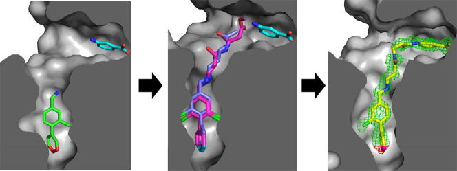
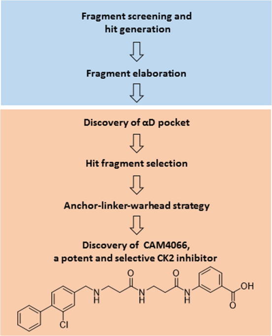
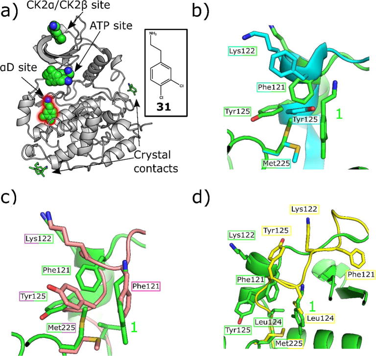
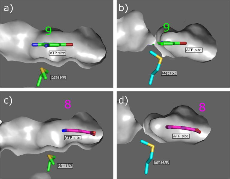
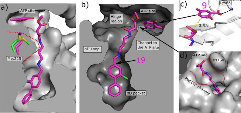
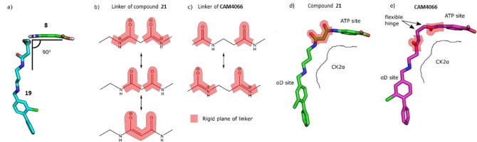
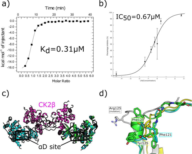
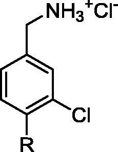
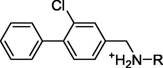
Recently we reported the discovery of a potent and selective CK2α inhibitor CAM4066. This compound inhibits CK2 activity by exploiting a pocket located outside the ATP binding site (αD pocket). Here we describe in detail the journey that led to the discovery of CAM4066 using the challenging fragment linking strategy. Specifically, we aimed to develop inhibitors by linking a high-affinity fragment anchored in the αD site to a weakly binding warhead fragment occupying the ATP site. Moreover, we describe the remarkable impact that molecular modelling had on the development of this novel chemical tool. The work described herein shows potential for the development of a novel class of CK2 inhibitors.

Keywords: CK2; Fragment linking; Fragment-based drug discovery; Kinase inhibition; Molecular modelling.


Figures

None
Graphical abstract
Scheme 1
Scheme 1
(a) DMF, RX, Na2CO3, (b) i) LiAlH4, Et2O, AlCl3, ii) Et2O, 2 M HCl in Et2O. General procedures are detailed in the Supporting information.
Scheme 2
Scheme 2
(a) CH2Cl2, Py, Tf2O, (b) ArB(OH)2, LiCl, DCE, Pd(PPh3)4, (c) i) LiAlH4, Et2O, AlCl3, ii) Et2O, 2 M HCl in Et2O.General procedures are detailed in the Supporting information.
Scheme 3
Scheme 3
(a) CH2Cl2, Py, Tf2O, (b) ArB(OH)2, LiCl, DCE, Pd(PPh3)4, (c) compounds 10, 1215: RNH2, DCE, NaBH(OAc)3, (d) compounds 11, 1618: RNH3+, MeOH, Et3N, NaBH(OAc)3, (e) Et2O, 2 M HCl in Et2O. General procedures are detailed in the Supporting information.
Scheme 4
Scheme 4
(a) CH2Cl2, 3-chloro-3-oxoproprionate, NaHCO3, (b) TFA/CH2Cl2, (c) MeOH, Et3N, NaBH(OAc)3, (d) LiOH,THF, 4 M HCl in dioxane, CH2Cl2. General procedures are detailed in the Supporting information.
Scheme 5
Scheme 5
(a) LiOH,THF, 4 M HCl in dioxane, CH2Cl2, (b) EDC-HCl, NMM, (c) RNH3+, MeOH, Et3N, NaBH(OAc)3, (d) LiOH,THF, 4 M HCl in dioxane.
Fig. 1
Fig. 1
Overview of the workflow from fragment screening against the α-β interface of CK2 to the discovery of a novel binding site and development of CAM4066.
Fig. 2
Fig. 2
a) Crystallographic structure of CK2α (grey) and 6 molecules of 1 (green). The promiscuous fragment occupies various sites of the protein showing potential for allosteric inhibitors (PDB code 5CLP). The molecules occupying biologically relevant sites are highlighted by sphere representation. The molecules at crystal contacts and therefore not of interest are shown as sticks. b) The movement of the important residues in the αD site upon the binding of 1 (PDB: 5CLP) compared to the partly open apo structure (PDB: 5CVH). c) The movement of the important residues in the αD site upon the binding of 1 (PDB: 5CLP) compared to the closed apo structure (PDB: 3FWQ) d) The movement of the important residues in the αD site upon the binding of 1 (PDB: 5CLP) compared to the inactive structure (PDB: 5CVG).
Fig. 3
Fig. 3
Schematic representation of the fragment elaboration carried out around 1 to develop a lead fragment to inhibit CK2.
Fig. 4
Fig. 4
The optimisation of the αD site fragment. a) The interactions of the amine of 1 with the backbone carbonyls of Val162 and Pro159 along with the interaction with Asn118 and Asn119 via a water bridge (PDB: 5CLP). b) The interactions of the amine of 7 with the backbone carbonyls of Val162 and Pro159 along with the interaction with Asn118 and Asn119 via a water bridge (PDB: 5CHS). Since the amine of 7 sits higher up in the pocket, it pulls down the top water into hydrogen bonding distance, thereby forming another water bridge to Asn118. c) The hydrophobic core of 1 sits in the hydrophobic pocket of the αD site (PDB: 5CLP), however there is still potential to optimise the interactions with this pocket. d) From the crystal structure it appears that 2 is more selective for the αD site over the ATP site, however, the OCF3 group does not fill the hydrophobic pocket of the αD site (PDB: 5CVF). e) The crystal structure of 7 bound in the αD site shows that the molecule fills the hydrophobic core of the αD pocket more efficiently (PDB: 5CHS). f) Movement of the αD loop upon binding of compounds 1 (green), 2 (magenta), 3 (cyan) and 4 (light blue).
Fig. 5
Fig. 5
Electron density of fragments 8 (PDB code 5CSP) and 9 (PDB code 5CSV) in the ATP binding site. The protein is represented in cyan and the H-bond between the fragments and the protein is shown as a black dashed line.
Fig. 6
Fig. 6
Modelling studies of potential linkers connecting fragments 7 and 8: a) A 9-atom linker needed to link the ATP site and the αD site. b) The proposed linker seen from above. Original fragments, as observed in crystal structures are highlighted with red and blue and the modelled linker if shows as semitransparent sticks.
Fig. 7
Fig. 7
Conformational changes of linker channel. (a) The crystal structure of 13 (PDB: 5CT0, pink) with 15 (PDB: 5CTP, light blue) superimposed. When 13 binds, Met163 does not move therefore the channel to the ATP site does not open and the surface of CK2α is clearly seen to block the binding of 15. The linker of 15 also forms a hydrogen bonding network with back bone carbonyls within the channel. b) The surface representation of the crystal structure of 13 (PDB: 5CT0) bound to CK2α. c) The surface representation of the crystal structure of 19 (PDB: CX9, purple) bound to CK2α with the structure of 13 (pink) superimposed. The outline of the surface when Met163 blocks the channel is represented by a red dotted line. d) Overlay of the modelled 7 atom (yellow) and 9 atom linkers on to the crystal structures of the fragments 7 (5CSH, light blue) and 8 (green) when Met163 has flipped to open the channel. The outline of the surface when Met163 blocks the channel is shown as a red dashed line; e) Overlay of the modelled 7 atom linker (yellow) and 9 atom linker (dark blue) when Met163 is flipped and forms the linker channel. The crystal structure of the ATP site binding fragment 8 is shown (green, 5CSP) The sides of the channel are highlighted by a red dashed line; f) Overlay of the modelled 7 atom linker (yellow) and 9 atom linker (dark blue) when Met163 has not flipped and blocks the linker channel. The crystal structure of the ATP site binding fragment 8 is shown (green, 5CSP).
Fig. 8
Fig. 8
a) The crystal structure of 9, a hinge binding fragment, bound to CK2α. Met163 (green) is in the down position where it blocks the channel between the αD site and the ATP site. b) The structure of 9 superimposed onto the structure of 19 where Met163 (blue) is flipped up and opens the channel between the αD site and the ATP site. Met163 would in this position clash with ZT0432. c) The crystal structure of 8 bound to CK2α. Met163 (green) is in the down position where it blocks the channel between the αD site and the ATP site. d) The structure of 8 superimposed onto the structure of 19 where Met163 (blue) is flipped up and opens the channel between the αD site and the ATP site. Met163 does not clash with 8.
Fig. 9
Fig. 9
Optimisation of the linker between the αD pocket and the ATP site. a) and b) The structure of 19 and 9 (PDB: 5CU2), co-soaked into CK2α. The surface of the channel when fragment 7 was bound in the αD pocket (PDB: 5CSH) is indicated by the red dashed line. The position of Met163 in the apo form is shown (green). c) A more detailed view of the structure of 9 co-soaked with 19, showing the location of the putative sites of linking (yellow dashed line); (d) The structure of 19 bound to CK2α showing the surface of the channel between the αD site and the ATP site formed by the movement of Met163.
Fig. 10
Fig. 10
The flexibility of the 2 linked inhibitors. a) The ATP site fragment 8 and the linker 19 are out of plane with each other by approximately 90° which makes successful linking more challenging. b) The various resonance forms of 21. The resonance forms impart rigidity to the linker in the position where flexibility is required. This may account for the reduced affinity. c) The various resonance forms of CAM4066. The resonance forms allow flexibility to the linker in the needed position. d) The binding conformation of 21 in CK2α. The highlighted areas are the rigid sections of the linker. e) The binding conformation of CAM4066 in CK2α. The highlighted areas are the rigid sections of the linker.
Fig. 11
Fig. 11
The inhibition of the CK2 holoenzyme by CAM4066. a) The isotherm of CAM4066 binding to the CK2 holoenzyme. b) The inhibition of kinase activity of the CK2 holoenzyme by CAM4066. c) The structure of the CK2 holoenzyme, CK2α is shown in blue and CK2β is shown in purple (PDB: 4MD7) with the structure of CAM4066 binding to CK2α, shown in green, (PDB: 5CU4) superimposed onto it. The position of the αD site is highlighted. d) A close up view of several conformations of the αD loop observed in the CK2 holoenzyme crystal structures with the structure of CAM4066 bound to CK2α, shown in green, (PDB: 5CU4) superimposed on them. The closed conformation is shown in light blue (PDB: 4MD7) the partly open form in yellow (PDB: 1 JWH) and the open conformation, in which Tyr125 is mutated to Arg125 is shown in grey (4DGL).
None
None
None

References

    1. Murray C.W., Rees D.C. Nat Chem. 2009;1:187–192. - PubMed
    1. Carr R.A.E., Congreve M., Murray C.W., Rees D.C. Drug Discovery Today. 2005;10:987–992. - PubMed
    1. Ciulli A., Abell C. Curr Opin Biotechnol. 2007;18:489–496. - PMC - PubMed
    1. Scott D.E., Coyne A.G., Hudson S.A., Abell C. Biochemistry. 2012;51:4990–5003. - PubMed
    1. Nazaré M., Matter H., Will D.W. Angew Chem Int Ed. 2012;51:905–911. - PubMed

MeSH terms