. 2017 Sep;31(9):3831-3847.
doi: 10.1096/fj.201601291R. Epub 2017 May 11.

PHD3 is a transcriptional coactivator of HIF-1α in nucleus pulposus cells independent of the PKM2-JMJD5 axis

Affiliations

PHD3 is a transcriptional coactivator of HIF-1α in nucleus pulposus cells independent of the PKM2-JMJD5 axis

Zachary R Schoepflin et al. FASEB J. 2017 Sep.

Abstract

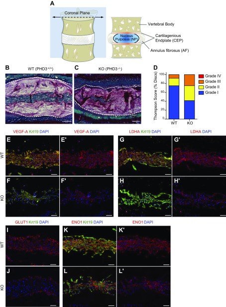
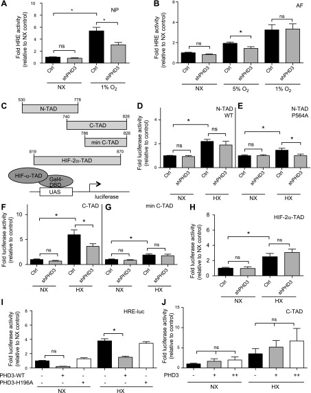
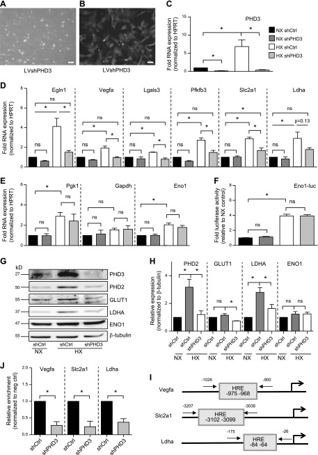
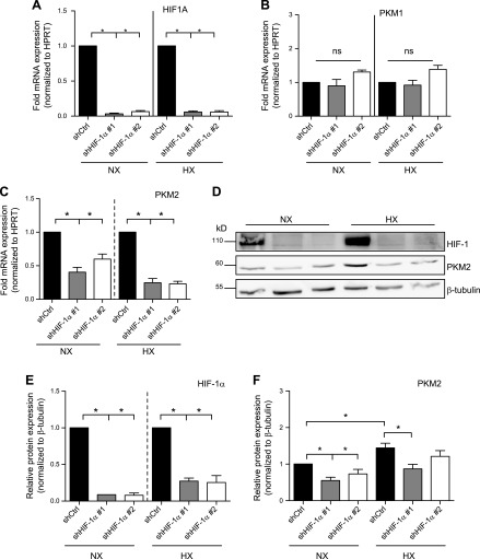
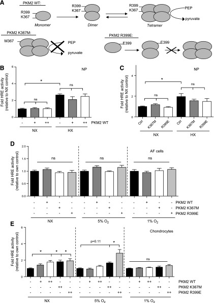
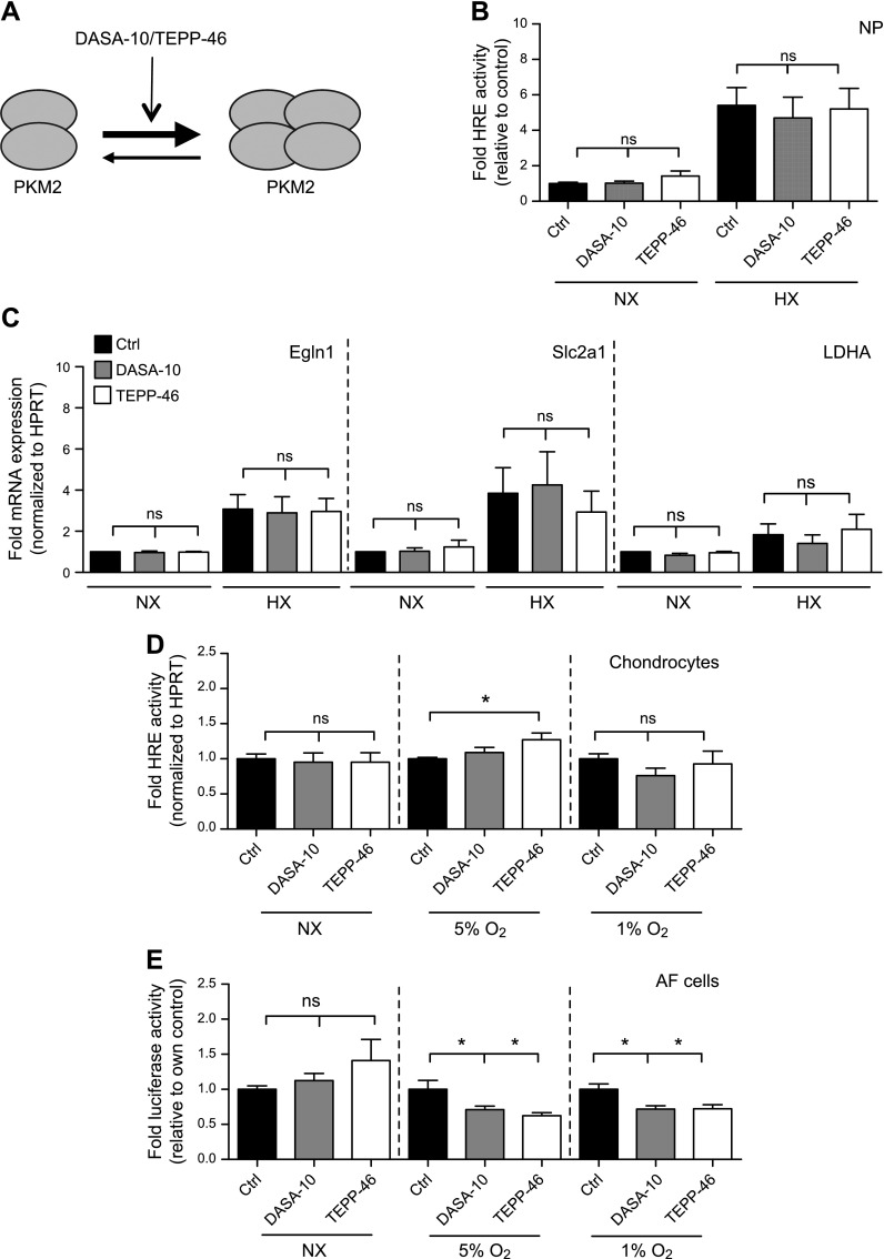
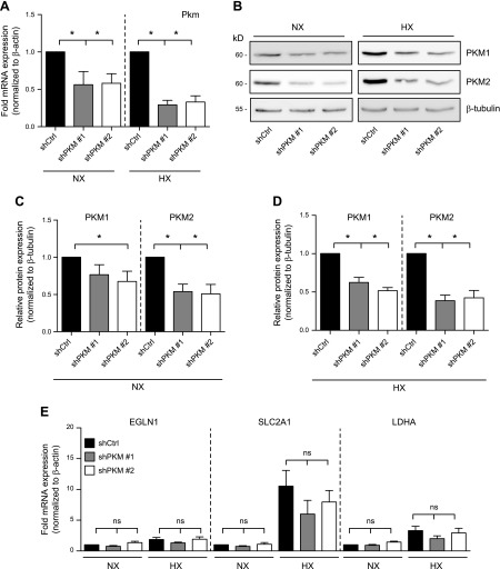
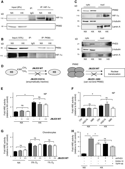
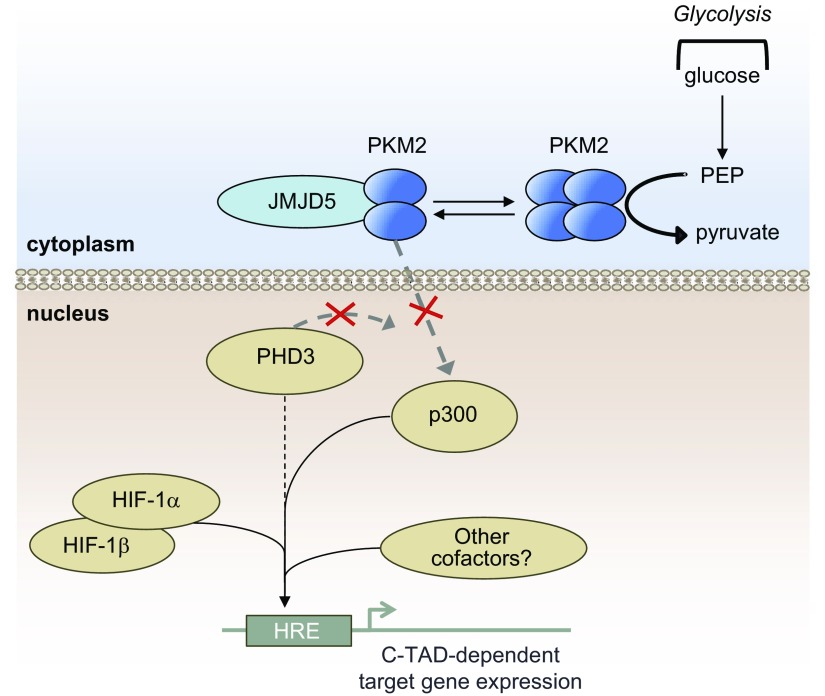
The role of prolyl hydroxylase (PHD)-3 as a hypoxia inducible factor (HIF)-1α cofactor is controversial and remains unknown in skeletal tissues. We investigated whether PHD3 controls HIF-1 transcriptional activity in nucleus pulposus (NP) cells through the pyruvate kinase muscle (PKM)-2-Jumonji domain--containing protein (JMJD5) axis. PHD3-/- mice (12.5 mo old) showed increased incidence of intervertebral disc degeneration with a concomitant decrease in expression of the HIF-1α targets VEGF-A, glucose transporter-1, and lactate dehydrogenase A. PHD3 silencing decreased hypoxic activation of HIF-1α C-terminal transactivation domain (C-TAD), but not HIF-1α-N-terminal-(N)-TAD or HIF-2α-TAD. Moreover, PHD3 suppression in NP cells resulted in decreased HIF-1α enrichment on target promoters and lower expression of select HIF-1 targets. Contrary to other cell types, manipulation of PKM2 and JMJD5 levels had no effect on HIF-1 activity in NP cells. Likewise, stabilization of tetrameric PKM2 by a chemical approach had no effect on PHD3-dependent HIF-1 activity. Coimmunoprecipitation assays showed lack of association between HIF-1α and PKM2 in NP cells. Results support the role of the PHD3 as a cofactor for HIF-1, independent of PKM2-JMJD5.-Schoepflin, Z. R., Silagi, E. S., Shapiro, I. M., Risbud, M. V. PHD3 is a transcriptional coactivator of HIF-1α in nucleus pulposus cells independent of the PKM2-JMJD5 axis.

Keywords: hypoxia; intervertebral disc; skeletal tissue; transcription factor.


Figures

Figure 1.
Figure 1.
PHD3−/− mice show increased incidence of disc degeneration and decreased expression of select HIF-1 target genes in NP tissue. A) Schematic drawing of spinal motion segment and coronal cross section showing vertebral bodies and the intervertebral disc with its central NP, circumferential AF, and superior and inferior cartilaginous endplates (CEP). B, C) Representative Safranin-O/Fast Green/hematoxylin–stained sections of 12.5-mo-old WT (B) and PHD3−/− (C) mice. The PHD3−/− mouse showed a decreased number of NP cells and some changes in cell morphology. D) PHD3−/− mice showed increased incidence of discs with a higher grade of degeneration, as assessed by the Thompson grading scale. Four discs/animal were scored from 3 pairs of littermate animals. E–L') Representative immunofluorescence images from 12.5-mo-old WT (PHD3+/+) and knockout (PHD3−/−) mice showing NP tissue areas with a comparable number of cells. There was decreased staining of HIF-1 targets VEGF-A (E–F'), LDHA (G–H'), and GLUT1 (I, J) in knockout mice compared to WT mice. In contrast, another HIF-1 target, ENO1, shows comparable expression in PHD3+/+ (K, K') and PHD3−/− (L, L') mice. Krt19 was used as a marker to label the NP tissue compartment with VEGF-A, LDHA, and ENO1 (independent channel not shown). Scale bars, 100 μm (B, C) and 50 μm (E–L'). Images are representative from 3 independent littermate groups.
Figure 2.
Figure 2.
PHD3 promotes hypoxic HIF-1α activity in disc cells through a C-TAD-dependent mechanism. A, B) Measurement of HIF activity in rat NP (A) (n = 7) and AF (B) (n = 3) cells using an HRE-luciferase reporter in NX and HX after PHD3 silencing. C) Gal4-HIF-α-TAD constructs used in panels D–H and the assay principle. D, E) Measurement of WTe (D) and P564A mutant (E) HIF-1α N-TAD (aa 530–778) activity in NP cells in NX and HX after PHD3 silencing (n = 3). F, G) Measurement of HIF-1α C-TAD (aa740–826) (F) (n = 7) and HIF-1α minimal C-TAD (aa 786–826) (G) (n = 3) activity in NP cells after PHD3 silencing. PHD3 silencing in HX resulted in decreased activity of HIF-1α C-TAD (aa 740–826) but not of minimal C-TAD (aa 786–826). H) Measurement of HIF-2α-TAD activity in NP cells after PHD3 silencing (n = 4). I) Measurement of HRE reporter activity in NP cells after overexpression of WT (PHD3-WT) or hydroxylase-deficient (PHD3-H196A) PHD3 (n = 3). J) PHD3 overexpression did not affect HIF-1α C-TAD activity (n = 3). Hypoxic culture for all experiments was 24 h. Data are means ± sem. Independent biologic experiments were performed with 3 technical replicates per experiment. *P < 0.05.
Figure 3.
Figure 3.
PHD3 controls expression of a select set of HIF-1 targets in NP cells through a p300-dependent mechanism. A, B) Bright-field (A) and fluorescent (B) images of rat NP cells after transduction with lentivirus coexpressing shPHD3 and enhanced green fluorescent protein. Scale bars, 10 μm. C) Measurement of PHD3 mRNA expression after transduction of NP cells with shRNA targeting PHD3 (n = 7). D) Measurement of mRNA expression of HIF-1 target genes in PHD3-silenced NP cells cultured in NX or HX for 72 h (n = 7). E) Hypoxic expression of Pgk1, Gapdh, and Eno1 in PHD3-silenced cells did not change (n = 3). F) Measurement of activity of Eno1 promoter containing 2 well-characterized HIF-1 binding sites after PHD3 knockdown (n = 7). G, H) Western blot (G) and corresponding densitometric analysis (H) of select HIF-1 targets in NP cells after stable knockdown of PHD3 (n = 4). I) Location of HRE sites within promoters of Vegfa, Slc2a1, and Ldha and location of ChIP primers used in J. J) HIF-1α enrichment at HRE sites within Vegfa, Slc2a1, and Ldha promoters decreases after knockdown of PHD3 during 72 h HX (n = 3). Luciferase assays were performed with 3 technical replicates per experiment; quantitative RT-PCR assays were performed with 2 technical replicates per experiment. Data are means ± sem. *P < 0.05.
Figure 4.
Figure 4.
Expression of the M2, but not M1, isoform of PK is induced by HIF-1 in NP cells. A) Measurement of HIF-1α mRNA after stable transduction of rat NP cells with 2 different lentivirally delivered shRNA sequences targeting HIF-1α (n = 4). B, C) mRNA expression of Pkm1 (B) and Pkm2 (C) in NP cells transduced with HIF-1α shRNAs (n = 3). DF) Western blot (D) and corresponding densitometric analysis of HIF-1α (E) and PKM2 (F) after stable knockdown of HIF-1α (n = 4). Data are means ± sem. *P < 0.05.
Figure 5.
Figure 5.
Overexpression of PKM2 does not affect HIF-1 activity in intervertebral disc cells, but does so in chondrocytes. A) WT, kinase-dead (K367M), and dimeric mutant (R399E) PKM2 constructs used in panels BE. B, C) HIF-1 activity in rat NP cells after overexpression of WT (B) or mutant (C) PKM2 did not change. D) HIF-1 activity in AF cells remained unaffected after overexpression of WT or mutant PKM2. E) HIF-1 activity in chondrocytes after overexpression of WT or mutant PKM2 shows increase in NX and at 5% O2, only with the R399E mutant. Hypoxic culture for all experiments was 24 h. Data are means ± sem of 3 independent experiments, performed with 3 technical replicates per experiment. *P < 0.05.
Figure 6.
Figure 6.
Stabilization of PKM2 tetramer using small molecular activators has no effect on HIF-1 activity in NP cells or chondrocytes. A) Mechanism of action of the small molecular activators DASA-10 and TEPP-46 used in panels B–E. B) HIF activity in rat NP cells after treatment with DASA-10 (20 μM) or TEPP-46 (50 μM) for 24 h (n = 3). C) mRNA expression of HIF-1 targets in NP cells showed no change after treatment with DASA-10 (20 μM) or TEPP-46 (50 μM) during NX and HX (n = 3). D) HIF-1 activity in chondrocytes did not decrease after stabilization of PKM2 tetramer by DASA-10 (20 μM) or TEPP-46 (50 μM) treatment for 24 h in 5 and 1% Po2 (n = 4). E) HIF-1 activity in rat AF cells after treatment with DASA-10 (20 μM) or TEPP-46 (50 μM) for 24 h decreases in 5 and 1% Po2 (n = 3). Luciferase assays were performed with 3 technical replicates per experiment. Data are means ± sem. *P < 0.05.
Figure 7.
Figure 7.
PKM2 silencing in NP cells does not control hypoxic expression of HIF-1 targets. A) Targeting of the Pkm locus in human NP cells by 2 independent lentivirally delivered shRNA sequences resulted in decreased Pkm mRNA expression (n = 5). BD) Western blot (B) and corresponding densitometric analysis (C, D) shows that both PKM1 and -2 levels are suppressed by either shRNA in NX (C) and HX (D) (n = 5). E) mRNA expression of HIF-1 target genes after PKM knockdown in NP cells (n = 5). Quantitative RT-PCR assays were performed with 2 technical replicates per experiment. Data are means ± sem. *P < 0.05.
Figure 8.
Figure 8.
PKM2 and HIF-1 do not interact in NP cells. A) Western blot analysis after immunoprecipitation of HIF-1α shows that HIF-1α does not interact with PKM2 in rat NP cells. Image is representative of 3 independent experiments. B) Western blot analysis after immunoprecipitation of PKM2 shows no evidence of PKM2-HIF-1α interaction in NP cells. Image is representative of 3 independent experiments. C) Western blot analysis of HIF-1α, PKM2, and PHD3 using cytoplasmic and nuclear fractions of NP cells. Images are representative of 3 independent experiments. D) Schematic of WT, demethylase deficient (H321A), and truncated (ΔN80, lacks PKM2 binding capability) JMJD5 constructs used in panels EG. E, F) HIF activity in NP cells after overexpression of WT (E) or mutant (F) JMJD5 constructs (n = 3). G) HIF activity in chondrocytes was also unaffected after overexpression of WT or mutant JMJD5 (n = 4). H) Measurement of HRE reporter activity in PHD3-silenced NP cells after pretreatment with PKM2 stabilizers (n = 3). Luciferase assays were performed with 3 technical replicates per experiment. Data are means ± sem. *P < 0.05.
Figure 9.
Figure 9.
PHD3 serves as a hypoxic transcriptional coactivator of HIF-1α C-TAD in NP cells in a p300-dependent and PKM2/JMJD5-independent manner. Proposed model of regulation of HIF-1 activity by PHD3 in NP cells. PHD3 serves as a hypoxic coactivator of HIF-1α in NP cells, directly or indirectly promoting HIF-1 enrichment at target loci and activating C-TAD-dependent targets, independent of PKM2/JMJD5. PKM2 in NP cells does not have cofactor function for HIF-1.

References

    1. Risbud M. V., Schipani E., Shapiro I. M. (2010) Hypoxic regulation of nucleus pulposus cell survival: from niche to notch. Am. J. Pathol. 176, 1577–1583 - PMC - PubMed
    1. U.S. Burden of Disease Collaborators (2013) The state of US health, 1990-2010: burden of diseases, injuries, and risk factors. JAMA 310, 591–608 - PMC - PubMed
    1. Scaal M. (2016) Early development of the vertebral column. Semin. Cell Dev. Biol. 49, 83–91 - PubMed
    1. Agrawal A., Guttapalli A., Narayan S., Albert T. J., Shapiro I. M., Risbud M. V. (2007) Normoxic stabilization of HIF-1alpha drives glycolytic metabolism and regulates aggrecan gene expression in nucleus pulposus cells of the rat intervertebral disk. Am. J. Physiol. Cell Physiol. 293, C621–C631 - PubMed
    1. Risbud M. V., Guttapalli A., Stokes D. G., Hawkins D., Danielson K. G., Schaer T. P., Albert T. J., Shapiro I. M. (2006) Nucleus pulposus cells express HIF-1 alpha under normoxic culture conditions: a metabolic adaptation to the intervertebral disc microenvironment. J. Cell. Biochem. 98, 152–159 - PubMed

Publication types

MeSH terms

LinkOut - more resources