. 2017 Jul 7;28(14):1950-1958.
doi: 10.1091/mbc.E17-01-0046. Epub 2017 May 11.

Mechanically patterned neuromuscular junctions-in-a-dish have improved functional maturation

Affiliations

Mechanically patterned neuromuscular junctions-in-a-dish have improved functional maturation

Cassandra L Happe et al. Mol Biol Cell. .

Abstract

Motor neuron (MN) diseases are progressive disorders resulting from degeneration of neuromuscular junctions (NMJs), which form the connection between MNs and muscle fibers. NMJ-in-a-dish models have been developed to examine human MN-associated dysfunction with disease; however such coculture models have randomly oriented myotubes with immature synapses that contract asynchronously. Mechanically patterned (MP) extracellular matrix with alternating soft and stiff stripes improves current NMJ-in-a-dish models by inducing both mouse and human myoblast durotaxis to stripes where they aligned, differentiated, and fused into patterned myotubes. Compared to conventional culture on rigid substrates or unpatterned hydrogels, MP substrates supported increased differentiation and fusion, significantly larger acetylcholine (ACh) receptor clusters, and increased expression of MuSK and Lrp4, two cell surface receptors required for NMJ formation. Robust contractions were observed when mouse myotubes were stimulated by ACh, with twitch duration and frequency most closely resembling those for mature muscle on MP substrates. Fused myotubes, when cocultured with MNs, were able to form even larger NMJs. Thus MP matrices produce more functionally active NMJs-in-a-dish, which could be used to elucidate disease pathology and facilitate drug discovery.


Figures

FIGURE 1:
FIGURE 1:
Fabrication of mechanically patterned hydrogel. (A) Patterning process by which sequential exposures of the hydrogel to UV light—the second exposure including a photomask—creates the patterned hydrogel. (B) Hydrogel pattern and plot of elastic modulus vs. position orthogonal to the pattern direction. Average modulus values for “soft” (teal) and “stiff” (blue) regions.
FIGURE 2:
FIGURE 2:
Durotactic pattern recognition and fusion marker expression. (A) Bright-field images of mouse myoblasts on the indicated substrates. Scale bar, 100 μm. Inset, low-magnification bright-field image of mouse myoblasts on the mechanically patterned substrate. Scale bar, 200 μm. (B) mRNA transcript levels for M-cadherin (left) and N-cadherin (right) for the indicated conditions. Data were normalized to glass substrates (n = 5).
FIGURE 3:
FIGURE 3:
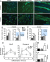
Improved myoblast fusion and differentiation on mechanically patterned matrices. (A) Representative fluorescence images of myotubes cultured on glass, myogenic (Myo), or mechanically patterned (MP) hydrogels at 5 d (mouse) or 7 d (human) after induction of differentiation. Cells were immunolabeled for myosin heavy chain (MHC, green) and counterstained for nuclei with Hoechst 33352 (blue). Scale bars, 30 μm. Fusion index (n = 10) and average myotube width (n = 30–88 individual myotubes per condition) for both (B) mouse and (C) human myotubes. The shaded region denotes the in vivo range of myotube width (Gaudel et al., 2008; Zhu et al., 2011; Lawlor et al., 2014; Gibbons et al., 2016). (D) mRNA expression of MyoD and myogenin normalized to day 0 for each condition plotted for the indicated number of days after induction of differentiation (n = 6). Data are shown for both substrates of myogenic stiffness, that is, 12 kPa (closed triangles), and with a mechanical pattern (open squares); expression levels are time dependent (p < 0.05). (E, F) MHC was assessed by qPCR, and data were normalized to GAPDH and plotted for glass, myogenic substrates, and mechanically patterned hydrogels (n = 5). *p < 0.05, **p < 0.01, and ***p < 0.001 based on ANOVA comparisons of the indicated groups.
FIGURE 4:
FIGURE 4:
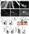
Patterning drives clustering of ACh receptors. (A) Representative fluorescence images of AChR clustering on agrin-treated myotubes cultured on glass, myogenic (Myo), or mechanically patterned (MP) hydrogels at 5 d (mouse) or 7 d (human) after induction of differentiation. AChRs were labeled with BTX. Scale bars, 25 μm. (B) Average AChR cluster size for mouse (left; n = 5) and human (right; n = 20). (C) Agrin-driven AChR clustering involving Lrp4 and MuSK. (D) Lrp4 and (E) MuSK expression was assessed by qPCR, and data were normalized to transcript expression on glass and plotted for the indicated species (n = 6). *p < 0.05 and ***p < 0.001 based on ANOVA comparisons of the indicated groups.
FIGURE 5:
FIGURE 5:
Patterning improves AChR clusters induced by MN coculture. (A) Representative fluorescence images of AChR clustering on mouse myotubes cocultured with mouse MN for 7 d. Myotubes were differentiated for 4 d before the start of the coculture. AChRs were labeled with BTX. Scale bar, 20 μm. Arrows indicate BTX immunoreactivity. (B) Average AChR cluster size (n = 20) for glass (Gl), soft myogenic substrates (Myo), and mechanically patterned hydrogels (MP). ***p < 0.001 based on ANOVA comparisons between substrate conditions. (C) Representative fluorescence image of patterned mouse myotube/mouse MN coculture at 7 d. Cells were immunostained with β-III-tubulin (TuJ1, green) and BTX (red) and counterstained for nuclei with Hoechst 33352 (blue). Scale bar, 30 μm. Arrows indicate BTX immunoreactivity that colocalizes with β-III-tubulin immunoreactivity, suggestive of a putative NMJ.
FIGURE 6:
FIGURE 6:
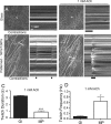
Functional assessment of AChR clusters in myotubes on different substrates. (A, B) Still movie images of myotube cultures (left). The white line indicates which contracting myotube was used to create the associated kymograph (right). The black bar at the bottom of each kymograph notes contractions. Cultures were stimulated with ACh as indicated. (C) Twitch duration and (D) frequency for glass (Gl; black) and mechanically patterned hydrogels (MP; white; n = 5). *p < 0.05 and ***p < 0.001 based on ANOVA comparisons between substrate conditions.
FIGURE 7:
FIGURE 7:
Substrate stiffness, but not myotube width, mediates AChR cluster formation on mechanically patterned substrates. (A) Elastic modulus of pathological mechanically patterned hydrogel. Fusion index (B) and myotube width (C) of mouse myotubes cultured on mechanically patterned hydrogels with myogenic (12 kPa) and pathological (58 kPa) stiff regions (n = 25). (D) Representative fluorescence images of AChR clustering on patterned hydrogels. AChR clusters are labeled with BTX. Scale bar, 10 μm. Arrows indicate AchR clusters. (E) AChR cluster area on myotubes cultured with agrin treatment on patterned hydrogels (n = 25). **p < 0.01 based on unpaired t test comparison. (F) Scatter plot of AChR cluster size vs. myotube width on mechanically patterned substrates with myogenic (12 kPa) stiff regions (n = 236 AChR clusters).

Similar articles

Cited by

References

    1. Abujarour R, Valamehr B. Generation of skeletal muscle cells from pluripotent stem cells: advances and challenges. Front Cell Dev Biol. 2015;3:29. - PMC - PubMed
    1. Altomare L, Riehle M, Gadegaard N, Tanzi MC, Fare S. Microcontact printing of fibronectin on a biodegradable polymeric surface for skeletal muscle cell orientation. Int J Artif Organs. 2010;33:535–543. - PubMed
    1. Bettadapur A, Suh GC, Geisse NA, Wang ER, Hua C, Huber HA, Viscio AA, Kim JY, Strickland JB, McCain ML. Prolonged culture of aligned skeletal myotubes on micromolded gelatin hydrogels. Sci Rep. 2016;6:28855. - PMC - PubMed
    1. Carlson BM. The biology of long-term denervated skeletal muscle. Eur J Transl Myol. 2014;24:3293. - PMC - PubMed
    1. Caron L, Kher D, Lee KL, McKernan R, Dumevska B, Hidalgo A, Li J, Yang H, Main H, Ferri G, et al. A human pluripotent stem cell model of facioscapulohumeral muscular dystrophy-affected skeletal muscles. Stem Cells Transl Med 5, 1145–1161. 2016 - PMC - PubMed

Substances