. 2017 Jun 5;216(6):1849-1864.
doi: 10.1083/jcb.201612030. Epub 2017 May 11.

Usher syndrome type 1-associated cadherins shape the photoreceptor outer segment

Affiliations

Usher syndrome type 1-associated cadherins shape the photoreceptor outer segment

Cataldo Schietroma et al. J Cell Biol. .

Abstract

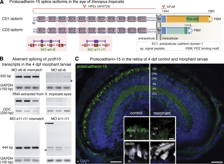
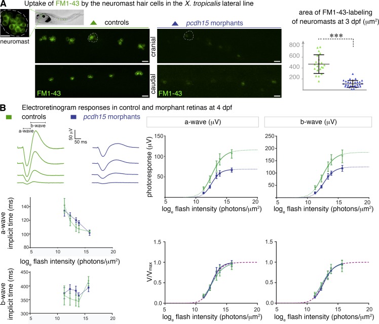
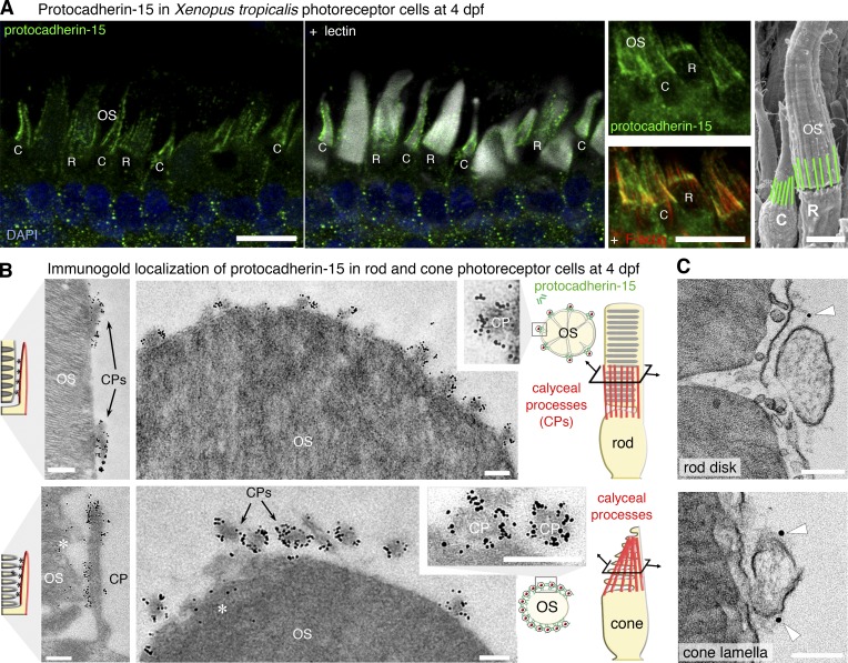
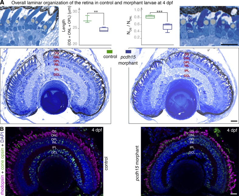
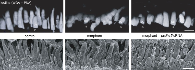
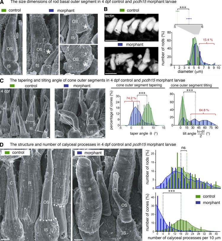
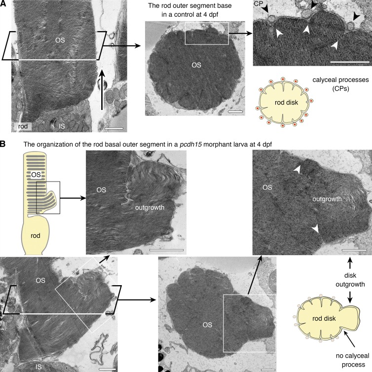
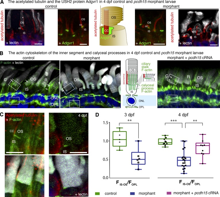
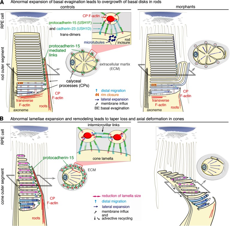
Usher syndrome type 1 (USH1) causes combined hearing and sight defects, but how mutations in USH1 genes lead to retinal dystrophy in patients remains elusive. The USH1 protein complex is associated with calyceal processes, which are microvilli of unknown function surrounding the base of the photoreceptor outer segment. We show that in Xenopus tropicalis, these processes are connected to the outer-segment membrane by links composed of protocadherin-15 (USH1F protein). Protocadherin-15 deficiency, obtained by a knockdown approach, leads to impaired photoreceptor function and abnormally shaped photoreceptor outer segments. Rod basal outer disks displayed excessive outgrowth, and cone outer segments were curved, with lamellae of heterogeneous sizes, defects also observed upon knockdown of Cdh23, encoding cadherin-23 (USH1D protein). The calyceal processes were virtually absent in cones and displayed markedly reduced F-actin content in rods, suggesting that protocadherin-15-containing links are essential for their development and/or maintenance. We propose that calyceal processes, together with their associated links, control the sizing of rod disks and cone lamellae throughout their daily renewal.


Figures

Figure 1.
Figure 1.
Antisense morpholino (MO) oligonucleotides block pcdh15 expression in the retina of X. tropicalis. (A) Predicted domain structure of the two splice isoforms of X. tropicalis protocadherin-15, positions of the antibodies used in the study, and pcdh15 MO e6-i6 and MO e11-i11. Aberrant splicing of pcdh15 transcripts is predicted to result in premature stop codons. (B) MO e6-i6 and MO e11-i11 affect pre-mRNA splicing and lead to several abnormal RT-PCR products (asterisks) because of intron retention, exon skipping, and the use of alternative splice sites. The overall reduction in the amount of the RT-PCR products is probably because of the nonsense-mediated decay of incorrectly spliced mRNAs. Abnormal transcripts and nonsense-mediated decay were not detected in control larvae microinjected with five-base mismatch control oligonucleotide. Ornithine decarboxylase (ODC) and glyceraldehyde 3-phosphate dehydrogenase (GAPDH) mRNAs were used as loading controls. (C) Lower levels of photoreceptor labeling for protocadherin-15 (green) in the 4 dpf larva microinjected with MO e6-i6 (right) than in the 4-dpf larva treated with the mismatch oligonucleotide control. Cell nuclei were stained with DAPI (blue). Insets: details of the inner segment (IS)–outer segment (OS) interface, with fluorescent lectins (white) and protocadherin-15 (green), staining is shown. ONL, outer nuclear layer; OPL, outer plexiform layer; INL, inner nuclear layer; IPL, inner plexiform layer; GCL, ganglion cell layer. Bars: (main) 20 µm; (insets) 5 µm.
Figure 2.
Figure 2.
Pcdh15 morphant larvae have altered mechanoelectrical transduction channels and photosensory deficits. (A) Uptake of FM1–43 dye by the hair cells of cranial and caudal neuromasts (high magnification of a neuromast, with 6–12 sensory hair cells shown). Dye uptake in neuromasts (some are outlined) is much weaker in 3-dpf pcdh15 morphants than it is in controls: 120.6 ± 15.5 µm2 in morphants (mean ± SEM; n = 33), versus 471.2 ± 37.5 µm2 in controls (n = 21; unpaired t test, ***, P < 0.0001). (B, top left) Representative electroretinogram traces (flash intensity increasing from bottom to top). (bottom left) The time-to-peak values obtained for the a- and b-waves (implicit times) did not differ significantly between controls and morphants. (right) Photoresponse curves (V, μV), plotted as a function of flash intensity and fitted with the Naka–Rushton function, in morphants and controls. The responses show a significant attenuation in morphants (comparison of fits by the least-squares method: P < 0.0001 for both waves), whereas after normalization by the maximum value Vmax, they do not differ significantly, giving similar values of I0.5 (P > 0.5 for both waves; bottom right panel; n = 7 controls, 13 morphants). Bars, 20 µm.
Figure 3.
Figure 3.
Protocadherin-15 is located at the calyceal processes of photoreceptor cells in X. tropicalis larvae. (A) In 4 dpf larvae, protocadherin-15 (green) is located around the base of the lectin-labeled (white) rod (R) and cone (C) outer segments (OS). Protocadherin-15 colocalizes with F-actin (red) that fill the calyceal processes (CPs). (B) In pre-embedding immunogold electron micrographs, silver-enhanced immunogold particles showed protocadherin-15 to be localized at the CPs surrounding the rod (top) and cone (bottom) OS. Sparse gold particles also decorate the lamellar membrane in cones (asterisks). (C) Protocadherin-15 immunolabeled gold particles (arrowheads) were localized with filaments connecting the CPs to the OS plasma membrane in rods (top) and cones (bottom). Bars: (A) 10 µm; (A, SEM image) 5 µm; (B) 200 nm; (C) 100 nm.
Figure 4.
Figure 4.
Pcdh15 knockdown in X. tropicalis does not affect retinal morphogenesis. (A) Semithin sections of control and morphant retinas at 4 dpf. The retinal layer shows similar organization in morphant and control retina. However, a loss of alignment and shape alterations of the photoreceptor outer segments (OS) are seen in the morphants (see high magnification of the boxed areas). The outer retina is significantly thinner in the morphants: thickness of the OS, outer nuclear layer (ONL), and outer plexiform layer (OPL) (L(OS + ONL + OPL) = 24.6 ± 0.3 µm, mean ± SEM, in morphants versus 27.7 ± 0.6 µm in controls, n = 3–4; unpaired t test, **, P = 0.005. The ratio of the number of OS profiles (NOS) to that of nuclei (NONL) is also significantly lower in morphants (0.55 ± 0.05, mean ± SEM, in morphants versus 0.83 ± 0.02 in controls, n = 5; unpaired t test, ****, P = 0.0005). (B) Cryosections of 4 dpf retinas stained with antibodies against rhodopsin (magenta) and cone opsin (green) show no evidence of opsin mislocalization in the morphant retina, but the shape and organization of both cones and rods are altered. INL, inner nuclear layer; IPL, inner plexiform layer; GCL, ganglion cell layer. Bars, 20 µm.
Figure 5.
Figure 5.
The photoreceptor outer segments are misaligned in pcdh15 morphant larvae. 3D rendering of the confocal stacks obtained from acrylamide-embedded vibratome sections (150 µm thick) stained with fluorescent lectin (top) and from SEM micrographs (bottom), highlighting the orderly arrangement of the subretinal space in 4 dpf control retinas (left). In contrast, age-matched morphant retinas contain photoreceptors with misshapen, curved, and misaligned outer segments (OS; middle). The coinjection into the embryo of cRNAs encoding the protocadherin-15 CD1 and CD3 isoforms with the splice-blocking morpholinos essentially preserved the well-ordered organization and parallel alignment of the OS in the larva (right). IS, inner segment, C, cone, R, rod. Bars, 10 µm.
Figure 6.
Figure 6.
Pcdh15 knockdown in X. tropicalis causes distinct morphological alterations to the rod and cone outer segments and the associated calyceal processes (scanning electron microscopy analyses). (A) In scanning electron micrographs, rod outer segments (OS) of 4-dpf morphants often display basal swelling and bulges, observed in 24 of 126 (19%) of morphant rods (n = 3 retinas, 95% CI, 17.3–19.3%). Occasionally, the base of the OS (asterisks) extended well beyond the edge of the inner segment (IS). Calyceal processes (CPs) are not detected adjacent to the basal outgrowths (asterisks). (B) In control retinas (top left), the basal diameters of the rod OS stained with WGA lectin (white) have a distribution close to normal (green bar graph and curve), centered on 5.1 ± 0.05 µm (mean ± SEM, n = 99; D’Agostino–Pearson test, P = 0.8; skewness: 0.15). In morphant retinas, the diameters have a non-normal distribution (blue bar graph and curve), centered on 5.5 ± 0.1 µm with a large positive skew (n = 104; D’Agostino–Pearson test, P < 0.0001; skewness: 1.16). In morphant retinas, the basal diameter of rods was significantly larger than in controls (unpaired t test, ***, P < 0.001), greater than two SDs from the control mean in 16 of 104 (15.4%) morphant rods. (C) At 4 dpf, control cone photoreceptors possess a slender OS coaxial with the IS and are characterized by a pronounced conical taper; both features are altered in the morphant cones. The OS tilt and taper were estimated on scanning electron micrographs by measuring the basal angles α and β and the taper angle θ at 5 µm from the IS–OS interface. The frequency histograms for tilt and taper angles show a narrow distribution of tilt angles centered on 5.8° (median value, n = 68) in control larvae, whereas in the morphants, the distribution is wider and flatter (median value, 36.5°; n = 54), reflecting considerable variability in tilt (K-S test, ***, P < 0.0001; bin width, 10°); 35 of 54 (64.8%) of the morphant cone OS had a tilt more than 2 SD greater than the control mean. The taper angle of the morphant cone OS was significantly smaller than that of the controls (2.7 ± 0.4°, n = 31; and 7.1 ± 0.3°, n = 32, respectively [mean ± SEM]; bin, 3°; K-S test, ***, P < 0.0001); 23 of 31 (74.2%) of morphant cone OS had a taper more than 2 SD less than the control mean. (D) We determined the number of calyceal processes (CPs) by counting them at their point of emergence (arrowheads) and plotted that number as linear density (number of CPs per 10 µm of IS–OS interface) for both rod (R) and cone (C) photoreceptors. Frequency distributions (bin width, 2 CPs per 10 µm) are significantly lower in morphant cones, but not in the rods. Mean densities in rods (in CPs per 10 µm): controls, 19.4 ± 1.6 (mean ± SEM, n = 24); morphants, 16.5 ± 1.5 (n = 33); K-S test, P = 0.27. Mean densities in cones: controls, 16.7 ± 0.8 (n = 53); morphants 3.7 ± 0.6 (n = 53); P < 0.0001. Bars: (A and C) 1 µm; (B) 10 µm; (D) 5 µm.
Figure 7.
Figure 7.
Pcdh15 knockdown in X. tropicalis causes distinct morphological alterations to the rod and cone outer segments and the associated calyceal processes (TEM analyses). (A) In longitudinal (left) and transverse (right) thin sections, control rod photoreceptors have the expected shape, with cylindrical, multilobed outer segments (OS). The calyceal processes (CPs) can be seen all around the OS, lying within (black arrowheads) and occasionally between incisures demarcating the lobes of the rod disks (see diagram). The base of the OS is congruent with the tip of the inner segment (IS; left, arrow). (B) In morphant larvae, several rods display an enlarged base (boxed areas), which TEM analysis revealed to consist of packets of outgrown disks extending well beyond the IS–OS interface and folding laterally. These outgrown disks are confined within the boundaries of the delimiting incisures (white arrowheads). The CPs are visible adjacent to the unaffected region but not adjacent to the basal enlargements of the OS (middle and right). Bars, 1 µm.
Figure 8.
Figure 8.
The actin cytoskeleton of the calyceal processes and their roots is disrupted in pcdh15 morphant larvae. (A) The axoneme, stained with antibodies against acetylated tubulin (red), is present in the morphant cone photoreceptors, but it is often curved and bent. The typical distribution of the USH2 protein Adgrv1 (green) at the base of the axoneme, in the periciliary ridge complex, is preserved in morphant photoreceptors. (B) At the inner segment (IS)–outer segment (OS) interface, there are two F-actin networks: the transverse F-actin network at the apical surface of the IS, and the longitudinal F-actin bundles of the calyceal processes and their roots (arrows). In 4-dpf morphant retinas, morphants display less phalloidin staining at the IS–OS interface (middle) than do the age-matched controls (left). F-actin staining in morphant photoreceptors is restored by the coinjection into the embryo of pcdh15 cRNAs with the pcdh15 MO e6-i6 (see arrows in the right panel). (C) The transverse F-actin network (green) can still be observed in a morphant photoreceptor. (D) Measurements of F-actin fluorescence intensity in the IS–OS interface (FIS–OS) compared with the subjacent region of the outer plexiform layer (OPL) (FOPL, taken as a reference) showed an FIS–OS/FOPL ratio that is 50% lower in the morphants than in the controls. The F-actin cytoskeleton was partially preserved in the retinas of 4-dpf morphants after the coinjection into the embryo of pcdh15 cRNAs and pcdh15 MO e6-i6 (FIS–OS/FOPL ratio 20% smaller than in controls). The FIS–OS/FOPL ratio at 3 dpf: controls, 1.05 ± 0.10 (n = 5); morphants, 0.54 ± 0.1 (n = 6), mean ± SEM; unpaired t test, **, P = 0.007. At 4 dpf: controls, 0.97 ± 0.03 (n = 10); morphants, 0.48 ± 0.06 (n = 20); morphants + cRNAs, 0.81 ± 0.08 (n = 9); unpaired t tests: controls versus morphants, ***, P < 0.0001; morphants versus morphants + cRNAs, **, P = 0.002; controls versus morphants + cRNAs, P = 0.05. Bars, 5 µm.
Figure 9.
Figure 9.
Model of the structural defects observed in the photoreceptors of morphant larvae. (A and B) In rod (A) and cone (B) photoreceptor cells, the basal evaginations (BEs) are formed with their leading edge exposed to the extracellular space. Calyceal processes (CPs) filled by F-actin (red) surround the base of the outer segment (OS) in control rods and cones (A and B, left). Links formed by trans-interactions between protocadherin-15 and cadherin-23 likely couple CPs to the OS membrane and to other CPs in the cones. The formation of trans-homotypic links, made up of either protocadherin-15 (Indzhykulian et al., 2013) or cadherin-23 cannot be excluded (A and B). (A, top left) Rod BEs form a continuous membrane system, topologically equivalent to cone lamellae. Lateral expansion of the BEs is tightly coupled to disk formation and distal displacement. Rim closure creates a discontinuity between the disk and the plasma membrane, precluding membrane redistribution in the plasmalemma. (top right) Uncontrolled lateral expansion of the nascent disks might lead to aberrant outgrowths, as shown in the pcdh15 morphant rods. In the morphant rods, the internal cytoskeleton of microtubules along the incisures may sustain the persisting CPs, although the F-actin cytoskeleton within the processes is markedly decreased. (B) Unlike rods, cones have no incisures, and the CPs do not anchor to microtubules. (bottom left) Preservation the cone OS shape requires a balanced distribution of forces and flows. Membrane input into the membrane folds of the cone lamellae at the OS base is equilibrated by advective recycling in the plasmalemma and apical shedding of older lamellae. This results in the lateral expansion, distal displacement, and progressive resizing of lamellae by retinal pigment epithelium (RPE) cells. (bottom right) In pcdh15 morphant cones lack CPs; disturbance of the balance between these antagonistic forces causes taper loss and an abnormal axial curvature of the OS.

Similar articles

Cited by

References

    1. Adato A., Michel V., Kikkawa Y., Reiners J., Alagramam K.N., Weil D., Yonekawa H., Wolfrum U., El-Amraoui A., and Petit C.. 2005. Interactions in the network of Usher syndrome type 1 proteins. Hum. Mol. Genet. 14:347–356. 10.1093/hmg/ddi031 - DOI - PubMed
    1. Ahmed Z.M., Riazuddin S., Bernstein S.L., Ahmed Z., Khan S., Griffith A.J., Morell R.J., Friedman T.B., Riazuddin S., and Wilcox E.R.. 2001. Mutations of the protocadherin gene PCDH15 cause Usher syndrome type 1F. Am. J. Hum. Genet. 69:25–34. 10.1086/321277 - DOI - PMC - PubMed
    1. Alagramam K.N., Yuan H., Kuehn M.H., Murcia C.L., Wayne S., Srisailpathy C.R.S., Lowry R.B., Knaus R., Van Laer L., Bernier F.P., et al. . 2001. Mutations in the novel protocadherin PCDH15 cause Usher syndrome type 1F. Hum. Mol. Genet. 10:1709–1718. (published erratum appears in Hum. Mol. Genet. 2001. 10:2603). 10.1093/hmg/10.16.1709 - DOI - PubMed
    1. Alagramam K.N., Miller N.D., Adappa N.D., Pitts D.R., Heaphy J.C., Yuan H., and Smith R.J.. 2007. Promoter, alternative splice forms, and genomic structure of protocadherin 15. Genomics. 90:482–492. 10.1016/j.ygeno.2007.06.007 - DOI - PMC - PubMed
    1. Bahloul A., Michel V., Hardelin J.P., Nouaille S., Hoos S., Houdusse A., England P., and Petit C.. 2010. Cadherin-23, myosin VIIa and harmonin, encoded by Usher syndrome type I genes, form a ternary complex and interact with membrane phospholipids. Hum. Mol. Genet. 19:3557–3565. 10.1093/hmg/ddq271 - DOI - PMC - PubMed

Publication types

MeSH terms

Associated data