. 2017 Jun 9;356(6342):1072-1076.
doi: 10.1126/science.aai8132. Epub 2017 May 11.

Macrophage function in tissue repair and remodeling requires IL-4 or IL-13 with apoptotic cells

Affiliations

Macrophage function in tissue repair and remodeling requires IL-4 or IL-13 with apoptotic cells

Lidia Bosurgi et al. Science. .

Abstract

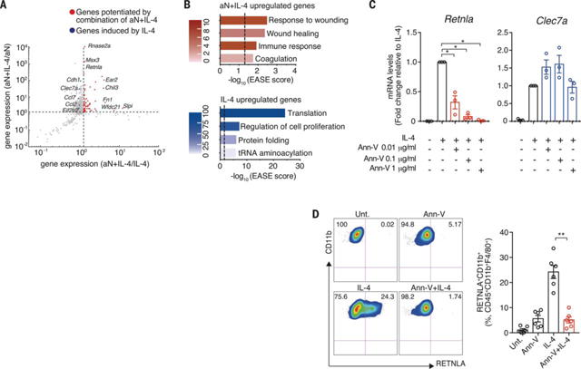
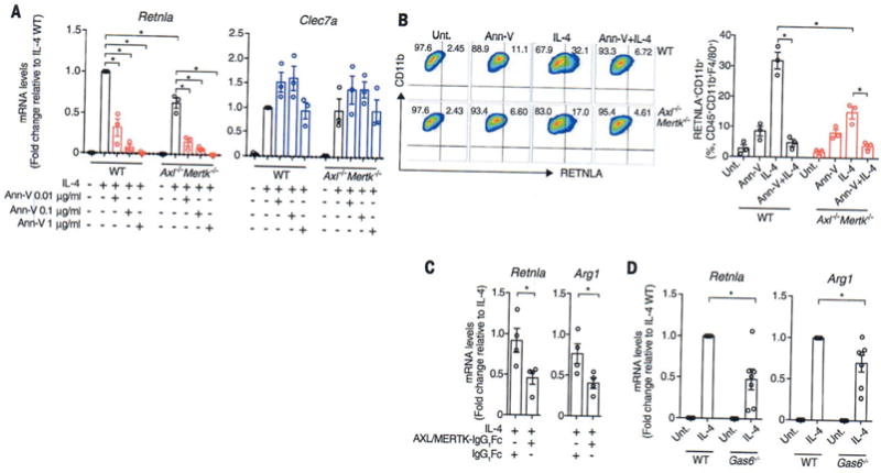
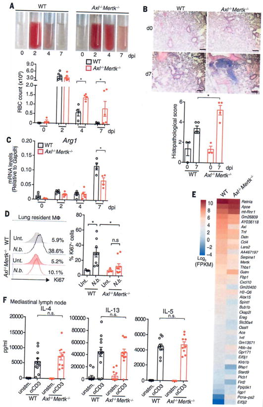
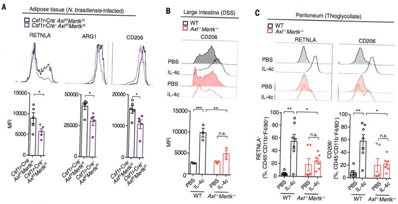
Tissue repair is a subset of a broad repertoire of interleukin-4 (IL-4)- and IL-13-dependent host responses during helminth infection. Here we show that IL-4 or IL-13 alone was not sufficient, but IL-4 or IL-13 together with apoptotic cells induced the tissue repair program in macrophages. Genetic ablation of sensors of apoptotic cells impaired the proliferation of tissue-resident macrophages and the induction of anti-inflammatory and tissue repair genes in the lungs after helminth infection or in the gut after induction of colitis. By contrast, the recognition of apoptotic cells was dispensable for cytokine-dependent induction of pattern recognition receptor, cell adhesion, or chemotaxis genes in macrophages. Detection of apoptotic cells can therefore spatially compartmentalize or prevent premature or ectopic activity of pleiotropic, soluble cytokines such as IL-4 or IL-13.


Figures

Fig. 1
Fig. 1. Codetection of IL-4 and apoptotic cells is required for the induction of an anti-inflammatory and tissue repair program in BMDMs
(A) Scatterplot of relative expression of genes induced in bone marrow-derived macrophages (BMDMs) upon exposure to apoptotic neutrophils (aN) or IL-4 or both, as determined by RNA-seq. Genes induced by aN, IL-4, or aN+JL-4 are shown in gray. Genes potentiated (>1.25-fold) by aN+IL-4 compared with both aN alone and IL-4 alone are shown in red. Representative genes induced by IL-4 but not potentiated by aN are shown in blue, n = 2 biological replicates. (B) Gene ontology assignments of genes potentiated in BMDMs exposed to aN+IL-4 or genes whose expression was induced in BMDMs treated with IL-4. Dashed lines represent P = 0.05. (C) mRNA expression normalized to Gapdh in BMDMs untreated or treated with indicated doses of annexin V (Ann-V) before stimulation with IL-4, as determined by qPCR. (D) Representative flow cytograms and independent data depicting frequency of RETNLA+ BMDMs untreated (Unt.) or treated with annexin V (1 μg/ml) before stimulation with IL-4 (Ann-V+IL-4). Each data point is from an independent sample. Mean ± SEM (error bars). *P < 0.05; **P < 0.01: Mann-Whitney U rank sum test.
Fig. 2
Fig. 2. IL-4 induction of anti-inflammatory and tissue repair genes in vitro depends on the activation of AXL and MERTK RTKs in BMDMs
(A) Expression of Retnla and Clec7a mRNA determined by qPCR and (B) representative flow cytograms and independent data of the frequency of RETNLA+ BMDMs from WT and Axl−/− Mertk−/− mice untreated (Unt.) or treated with annexin V at indicated doses (A) or 1 μg/ml (B) before stimulation with IL-4. (C) Expression of Retnla and Arg1 mRNA determined by qPCR in WT BMDMs incubated with IL-4 in the presence of AXL and MERTK extracellular domain-Fc chimeric proteins or IgGrFc control (IgG, immunoglobulin G). (D) Expression of Retnla and Arg1 mRNA determined by qPCR in BMDMs from WT and Gas6−/− mice upon stimulation with IL-4. Gapdh expression was used for normalization. Each data point is from an independent sample. Mean ± SEM (error bars). *P < 0.05; Mann-Whitney U rank sum test.
Fig. 3
Fig. 3. IL-4 and/or IL-13 induction of anti-inflammatory and tissue repair responses in macrophages upon infection with N. brasiliensis is dependent on AXL and MERTK RTKs
WT and Axl−/− Mertk−/− mice were infected with N. brasiliensis. (A) Representative images and independent data showing red blood cell (RBC) counts at the indicated number of days postinfection (dpi) in the bronchoalveolar lavage. (B) Representative images and histopathological score of lung sections stained with hematoxylin and eosin. Scale bars, 5 μm. (C) Arg1 expression in the lungs, as detected by qPCR. (D) Row cytometric analysis showing the frequency of Ki67+ lung macrophages (Mφ) (CD45+CD11chiSiglecF+CD11blo) from WT and Axl−/− Mertik−/− mice, untreated (Unt.) or 9 dpi. N.b., N. brasiliensis. (E) Heat map representing the most differentially expressed genes [log2 of expression levels, fragments per kilobase of transcript per million mapped reads (FPKM)] in lung macrophages from WT and Axl−/− Mertk−/− mice at 7 dpi, as determined by RNA-seq. (F) IL-4, IL-13, and IL-5 production by T cells from WT and Axl−/− Mertk−/− mice at 7 dpi. unstm., unstimulated. Each data point is from an independent sample. For (A) and (B), one experiment, which is representative of three independent experiments, is reported. Mean ± SEM (error bars). n.s., nonsignificant; *P < 0.05; Mann-Whitney U rank sum test.
Fig. 4
Fig. 4. IL-4 and AXL or MERTK RTK codetection functions across multiple tissues to induce anti-inflammatory and tissue repair-associated genes in macrophages
(A) Macrophages (CD45+CD11b+F4/80+) isolated from the visceral adipose tissue of Csf1r-CreAxlf/fMertkf/f and Csf1r-Cre+Axlf/fMertkf/f animals at 7 dpi with N. brasiliensis were analyzed for expression of RETNLA, ARG1, and CD206 by flow cytometry. Histograms and independent data, representative of two independent experiments, are reported. MFI, mean fluorescence intensity. (B) Expression of CD206 on lamina propria macrophages (CD45+CD11b+F4/80hl) isolated from the large intestine of WT and Axl−/−Mertk−/− mice treated for 7 days with 1.5% DSS and injected on days 3 and 5 with IL-4c or PBS intraperitoneally. Histograms and independent data are reported. (C) Frequency of RETNLA+ and CD206+ CD45+CD11b+F4/80+ peritoneal macrophages from WT and Axl−/−Mertk−/− mice injected intraperitoneally with 4% thioglycollate on day 0, followed by intraperitoneal injection of IL-4c or PBS on day 2 and isolation on day 4. Histograms and independent data, representative of two independent experiments, are shown. Mean ± SEM (error bars). n.s., nonsignificant; *P < 0.05; **P < 0.01; ***P < 0.001; Mann-Whitney U rank sum test.

Comment in

Similar articles

Cited by

References

    1. Murray PJ, et al. Immunity. 2014;41:14–20. - PMC - PubMed
    1. Mantovani A, Biswas SK, Galdiero MR, Sica A, Locati M. J Pathol. 2013;229:176–185. - PubMed
    1. Van Dyken SJ, Locksley RM. Annu Rev Immunol. 2013;31:317–343. - PMC - PubMed
    1. Gordon S, Martinez FO. Immunity. 2010;32:593–604. - PubMed
    1. Poon IK, Lucas CD, Rossi AG, Ravichandran KS. Nat Rev Immunol. 2014;14:166–180. - PMC - PubMed

Publication types