. 2017 Jun 9;356(6342):1076-1080.
doi: 10.1126/science.aaj2067. Epub 2017 May 11.

Local amplifiers of IL-4Rα-mediated macrophage activation promote repair in lung and liver

Affiliations

Local amplifiers of IL-4Rα-mediated macrophage activation promote repair in lung and liver

Carlos M Minutti et al. Science. .

Abstract

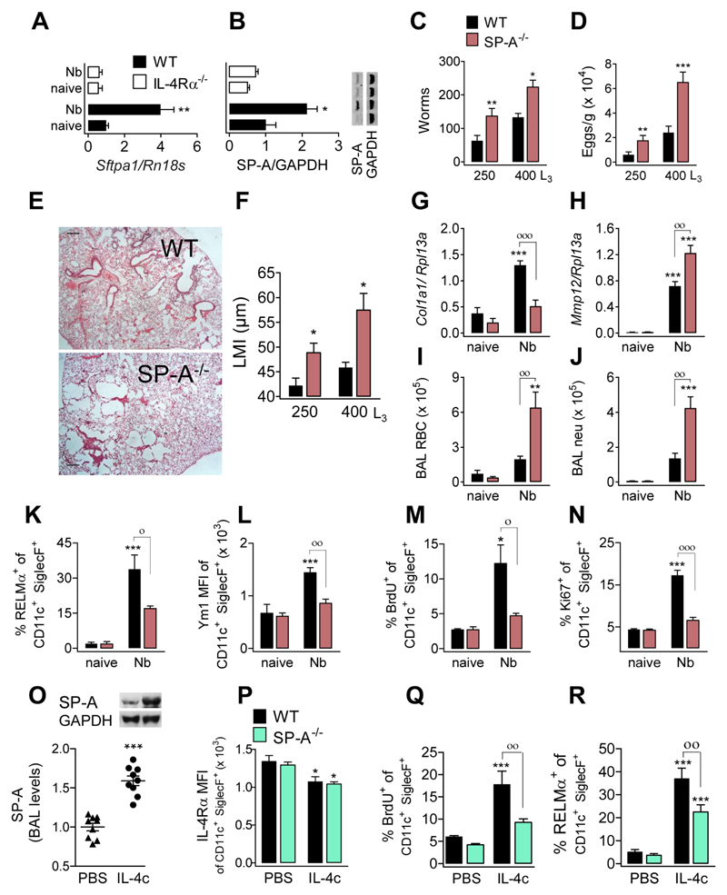
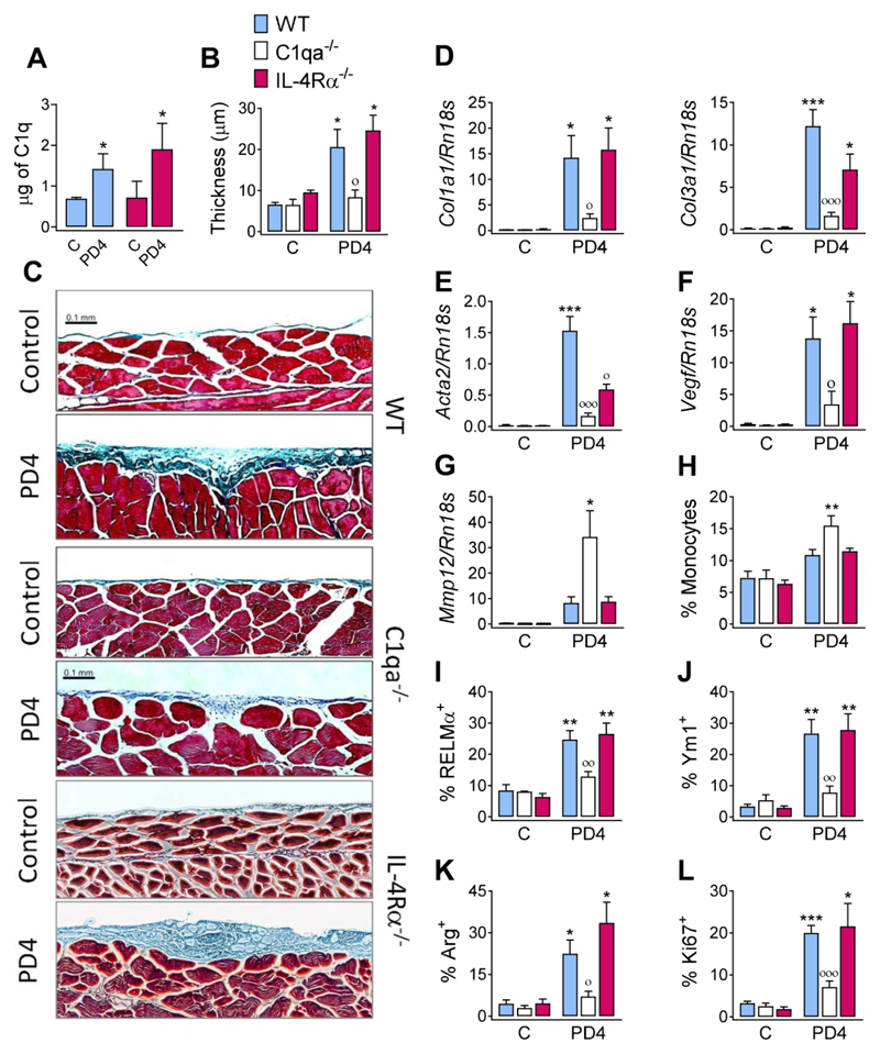
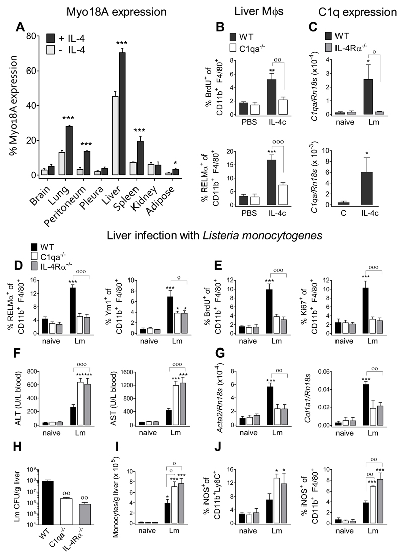
The type 2 immune response controls helminth infection and maintains tissue homeostasis but can lead to allergy and fibrosis if not adequately regulated. We have discovered local tissue-specific amplifiers of type 2-mediated macrophage activation. In the lung, surfactant protein A (SP-A) enhanced interleukin-4 (IL-4)-dependent macrophage proliferation and activation, accelerating parasite clearance and reducing pulmonary injury after infection with a lung-migrating helminth. In the peritoneal cavity and liver, C1q enhancement of type 2 macrophage activation was required for liver repair after bacterial infection, but resulted in fibrosis after peritoneal dialysis. IL-4 drives production of these structurally related defense collagens, SP-A and C1q, and the expression of their receptor, myosin 18A. These findings reveal the existence within different tissues of an amplification system needed for local type 2 responses.


Conflict of interest statement

Competing financial interests

The authors declare no conflict of interest

Figures

Fig. 1
Fig. 1. Higher worm burden, greater nematode-induced lung damage and reduced IL-4-induced proliferation and activation in mice lacking SP-A.
Samples were assessed at d6 after N. brasiliensis infection. SP-A (A) mRNA and (B) protein expression in lung tissue of WT and IL-4Rα-/- mice. (C) Adult larvae in the small intestine, (D) egg output in faeces, and (E) microscopy of H&E stained lung sections (scale bars, 500 μm) in WT and SP-A-/- mice. (F) Lung damage, quantified by ‘mean linear intercept’ from micrographs of H&E stained lung sections. (G) Amplification of Col1a1- and (H) Mmp12-encoding mRNA in lung tissue. Number of (I) red blood cells and (J) neutrophils isolated in BAL. Expression of (K) RELMα and (L) Ym1 by aMϕ from BAL. Because all aMϕ are Ym1 positive, MFI is shown for Ym1. (M) BrdU incorporation and (N) Ki67 expression by aMϕ from BAL. Data are representative from two independent experiments (mean ± SEM; naïve: 3 mice, Nb: 6 mice). (O-R) WT and SP-A-/- mice treated with 5μg IL-4c (i.p.) at days 0 and 2 and analyzed at day 4. (O) Relative SP-A levels in BAL (representative western blot shown) of WT mice treated with IL-4c or PBS. (P) Expression of IL-4Rα by aMϕ from BAL. (Q) BrdU incorporation and (R) RELMα expression in aMϕ. Data pooled from three independent experiments (means ± SEM) (PBS: 9 mice, IL-4c: 11 mice). ANOVA followed by the Bonferroni multiple-comparison test was used. *p < 0.05, **p < 0.01, and ***p < 0.001 when compared with the untreated/uninfected group. °p < 0.05, °°p < 0.01 and °°°p < 0.001 when WT vs. SP-A-/- groups are compared.
Fig. 2
Fig. 2. SP-A and C1q act through Myo18A to enhance IL-4-induced proliferation and activation of alveolar and peritoneal macrophages, respectively.
(A) Murine macrophages were treated with IL-4 in the presence or absence of SP-A or C1q. BrdU incorporation and secretion of Ym1 are shown. (B) For aMϕs, 5 μg IL-4c was delivered ip at d0 and d2, and BAL cells analyzed at d4. For pMϕs, 1 μg IL-4c was delivered ip at d0, and peritoneal cells analyzed at d1: BrdU incorporation and RELMα expression are shown. (C) Murine macrophages were treated with anti-Myo18A or rabbit IgG plus either IL-4+SP-A (aMϕ) or IL-4+C1q (pMϕ). BrdU incorporation and secretion of Ym1 are shown. (D) Myo18A expression on the surface of macrophages from WT, SP-A-/-, and C1qa-/- mice treated with or without IL-4c as described in (B). Concurrently with IL-4c delivery, some WT mice were intra-nasally treated with either anti-Myo18A or rabbit IgG antibody at d2 and d3, and samples analyzed at d4. BrdU incorporation is shown. All statistical analysis was performed by ANOVA followed by the Bonferroni multiple-comparison test. (A+C) Results are presented as means (± SEM) from three different cell cultures with at least three biological replicates. *p < 0.05, **p < 0.01, and ***p < 0.001, when compared with untreated cells; °p < 0.05, °°p < 0.01, and °°°p < 0.001, when SP-A+IL-4- or C1q+IL4-treated are compared with IL-4-treated; ##p < 0.01, and ###p < 0.001, the effect of anti-Myo18A antibody on cells treated with SP-A+IL-4 or C1q+IL4. (B+D) Data were pooled from three independent experiments (means ± SEM) (PBS: 6 mice, other groups: 9 mice). *p < 0.05, **p < 0.01, and ***p < 0.001, when compared with PBS treated mice; °p < 0.05, when WT vs. C1qa-/- mice treated with IL-4c are compared (B) °p < 0.05, °°p < 0.01, and °°°p < 0.001 when anti-Myo18A vs rabbit IgG treatment is compared in IL-4c-treated mice (D).
Fig. 3
Fig. 3. C1q enhances peritoneal fibrosis induced by a lactate dialysate.
WT, C1qa-/- or IL-4Rα-/- mice were either untreated (C) or treated with a 14 ip injections of Dianeal PD-4 every other day. Samples were analyzed a day after the last delivery. (A) Total amount of C1q in the peritoneal washes was determined by ELISA. (B) Quantification of the thickness of the submesothelial compact zone from (C) microscopy of Masson’s trichrome stained parietal peritoneum slices (scale bars, 0.1 mm). Amplification of (D) Col1a1, Col3a1, (E) Acta2, (F) Vegf, and (G) Mmp12-encoding mRNA in peritoneal tissue. (H) Percentage of infiltrating monocytes as quantified by FACS. Expression of (I) RELMα, (J) Ym1, (K) Arg and (L) Ki67 by peritoneal macrophages. Results are representative from two independent experiments (means ± SEM) (untreated: 3 mice, PD4: 6 mice). ANOVA followed by the Bonferroni multiple-comparison test or Student’s t-test (A) was used. *p < 0.05, **p < 0.01, and ***p < 0.001 when compared with control group; °p < 0.05, °°p < 0.01, and °°°p < 0.001 when WT vs. C1qa-/- mice treated with Dianeal PD-4 are compared.
Fig. 4
Fig. 4. C1q is required for appropriate macrophage activation in the liver during Listeria monocytogenes infection.
(A, B, C) WT or C1qa-/- mice received 1 μg IL-4c (ip) at d0, and samples were analyzed at d1. (A) Myo18A expression on the surface of resident macrophages from the indicated tissues. Resident macrophages were identified as described in the methods (B) BrdU incorporation and RELMα expression of liver macrophages. (C) (lower panel) IL-4-induced amplification of C1q-encoding mRNA in the liver. (C-J) WT, C1qa-/-, or IL-4Rα-/- mice were left uninfected or received intravenous infection with 104 L. monocytogenes c.f.u. and samples were assessed at 3.5 dpi. (C) (upper panel) Lm-induced amplification of C1q-encoding mRNA. (D) Expression of RELMα and Ym1 by liver macrophages. (E) BrdU incorporation and Ki67 expression by liver macrophages. (F) Quantification of ALT and AST in serum. (G) Amplification of Acta2- and Col1a1-encoding mRNA in the liver. (H) Liver bacterial load. (I) Number of monocytes in liver single cell suspensions. (J) iNOS expression by liver monocytes and macrophages. Data are representative from two independent experiments (mean ± SEM; naïve: 4 mice, Lm: 5 mice). ANOVA followed by the Bonferroni multiple-comparison test was used. *p < 0.05, and ***p < 0.001, when compared with the uninfected group; °p < 0.05, °°p < 0.01, and °°°p < 0.001 when WT vs. C1qa-/- or IL-4Rα-/- infected groups are compared.

Comment in

Similar articles

Cited by

References

    1. Murray PJ, et al. Macrophage activation and polarization: nomenclature and experimental guidelines. Immunity. 2014;41:14–20. - PMC - PubMed
    1. Grencis RK. Immunity to helminths: resistance, regulation, and susceptibility to gastrointestinal nematodes. Annual review of immunology. 2015;33:201–225. - PubMed
    1. Chen F, et al. An essential role for TH2-type responses in limiting acute tissue damage during experimental helminth infection. Nature medicine. 2012;18:260–266. - PMC - PubMed
    1. Wynn TA, Vannella KM. Macrophages in Tissue Repair, Regeneration, and Fibrosis. Immunity. 2016;44:450–462. - PMC - PubMed
    1. Wright JR. Immunoregulatory functions of surfactant proteins. Nature reviews Immunology. 2005;5:58–68. - PubMed

Publication types