. 2017 Jul 7;292(27):11189-11205.
doi: 10.1074/jbc.M117.782326. Epub 2017 May 11.

Endoplasmic reticulum (ER) Ca2+-channel activity contributes to ER stress and cone death in cyclic nucleotide-gated channel deficiency

Affiliations

Endoplasmic reticulum (ER) Ca2+-channel activity contributes to ER stress and cone death in cyclic nucleotide-gated channel deficiency

Michael R Butler et al. J Biol Chem. .

Abstract

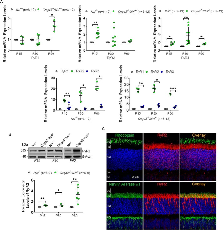
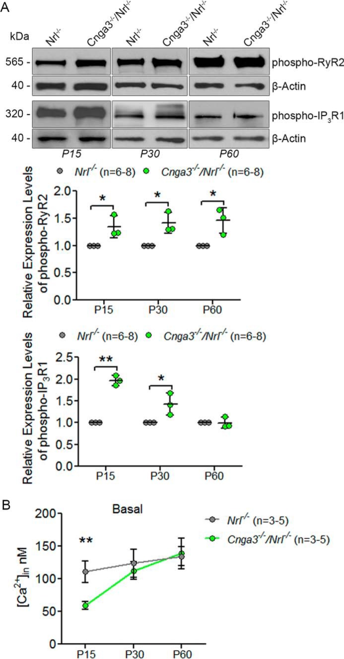
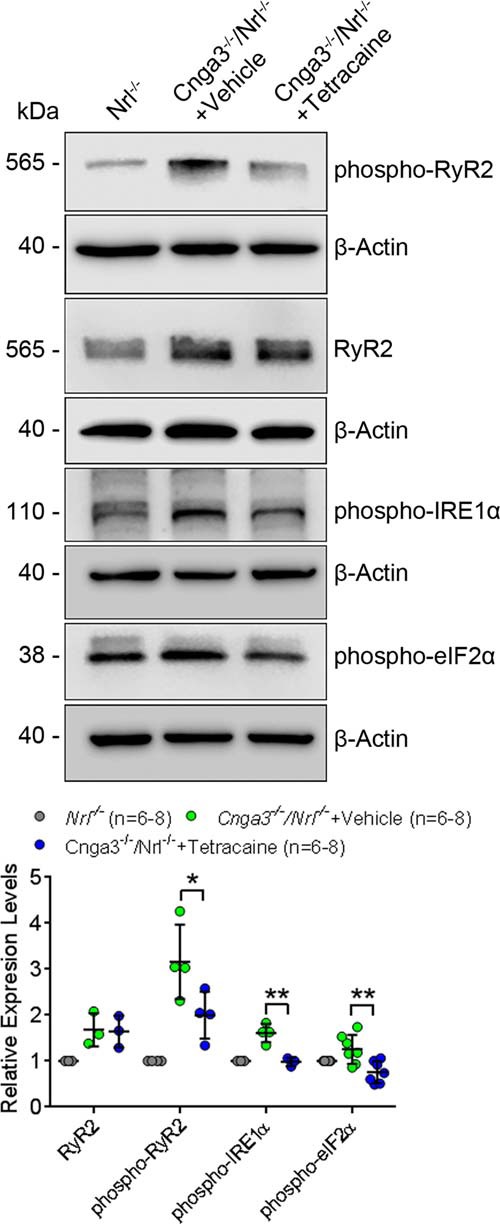
Endoplasmic reticulum (ER) stress and mislocalization of improperly folded proteins have been shown to contribute to photoreceptor death in models of inherited retinal degenerative diseases. In particular, mice with cone cyclic nucleotide-gated (CNG) channel deficiency, a model for achromatopsia, display both early-onset ER stress and opsin mistrafficking. By 2 weeks of age, these mice show elevated signaling from all three arms of the ER-stress pathway, and by 1 month, cone opsin is improperly distributed away from its normal outer segment location to other retinal layers. This work investigated the role of Ca2+-release channels in ER stress, protein mislocalization, and cone death in a mouse model of CNG-channel deficiency. We examined whether preservation of luminal Ca2+ stores through pharmacological and genetic suppression of ER Ca2+ efflux protects cones by attenuating ER stress. We demonstrated that the inhibition of ER Ca2+-efflux channels reduced all three arms of ER-stress signaling while improving opsin trafficking to cone outer segments and decreasing cone death by 20-35%. Cone-specific gene deletion of the inositol-1,4,5-trisphosphate receptor type I (IP3R1) also significantly increased cone density in the CNG-channel-deficient mice, suggesting that IP3R1 signaling contributes to Ca2+ homeostasis and cone survival. Consistent with the important contribution of organellar Ca2+ signaling in this achromatopsia mouse model, significant differences in dynamic intraorganellar Ca2+ levels were detected in CNG-channel-deficient cones. These results thus identify a novel molecular link between Ca2+ homeostasis and cone degeneration, thereby revealing novel therapeutic targets to preserve cones in inherited retinal degenerative diseases.

Keywords: CNG channel; calcium channel; endoplasmic reticulum stress (ER stress); inositol trisphosphate receptor (InsP3R); photoreceptor; ryanodine receptor.


Conflict of interest statement

The authors declare that they have no conflicts of interest with the contents of this article

Figures

Figure 1.
Figure 1.
Expression of the IP3R1 isoform is increased in Cnga3−/−;Nrl−/− mice and is localized to the inner segment in cones. A, retinas prepared from Cnga3−/−;Nrl−/− and Nrl−/− mice were analyzed for expression of IP3R isoforms by qRT-PCR at P15, P30, and P60 following normalization to Hprt1 mRNA controls. B, total IP3R1 isoform protein levels were measured at P15, P30, and P60 in Cnga3−/−;Nrl−/− and Nrl−/− mice. Shown are representative Western blot images with corresponding densitometric analysis following normalization to internal loading control β-actin. C, localization of the IP3R1 isoform was determined via immunofluorescence co-labeling with rhodopsin or Na+/K+ ATPase α1 subunit at P30 in wild-type mice. ONL, outer nuclear layer; OPL, outer plexiform layer; INL, inner nuclear layer. Data are presented as mean ± S.D. of at least three independent assays using retinas from at least six mice/group. Unpaired Student's t test was used to determine significance between CNG-channel-deficient and Nrl−/− mice (*, p < 0.05; **, p < 0.01; ***, p < 0.001).
Figure 2.
Figure 2.
Expression of the RyR2 isoform is increased in Cnga3−/−;Nrl−/− mice and is localized to the inner segment in cones. A, retinas prepared from Cnga3−/−;Nrl−/− and Nrl−/− mice were analyzed for expression of RyR isoforms by qRT-PCR at P15, P30, and P60 following normalization to Hprt1 mRNA controls. B, total RyR2 isoform protein levels were measured at P15, P30, and P60 in Cnga3−/−;Nrl−/− and Nrl−/− mice. Shown are representative Western blot images with corresponding densitometric analysis following normalization to internal loading control β-actin. C, localization of the RyR2 isoform was determined via immunofluorescence co-labeling with rhodopsin or Na+/K+ ATPase α1 subunit at P30 in wild-type mice. ONL, outer nuclear layer; OPL, outer plexiform layer; INL, inner nuclear layer. Data are presented as mean ± S.D. of at least three independent assays using retinas from at least six mice/group. Unpaired Student's t test was used to determine significance between CNG-channel-deficient and Nrl−/− mice (*, p < 0.05; **, p < 0.01; ***, p < 0.001).
Figure 3.
Figure 3.
Expression of phospho-IP3R1 and phospho-RyR2 is increased and intracellular Ca2+ is decreased at early ages in Cnga3−/−;Nrl−/− mice. A, retinal protein expression levels of phospho-RyR2 and phospho-IP3R1 at P15, P30, and P60 in Cnga3−/−;Nrl−/− and Nrl−/− mice were analyzed. Shown are representative Western blot images with corresponding densitometric analysis following normalization to internal loading control β-actin. B, basal intracellular Ca2+levels were also measured in Cnga3−/−;Nrl−/− and Nrl−/− mice at P15, P30, and P60. Data are presented as mean ± S.D. of at least three independent assays using retinas from 6–8 mice/group for immunoblotting and 15–33 photoreceptors from 3–5 mice for [Ca2+]i assays. Unpaired Student's t test was used to determine significance between CNG-channel-deficient and Nrl−/− mice (*, p < 0.05; **, p < 0.01).
Figure 4.
Figure 4.
Expression of ER-chaperone proteins is increased in Cnga3−/−;Nrl−/− mice and luminal Ca2+ stores undergo dynamic changes. A, retinas prepared from Cnga3−/−;Nrl−/− and Nrl−/− mice were analyzed for protein expression of Grp78/BiP and calreticulin by Western blot analysis at P15, P30, and P60. Shown are representative Western blot images with corresponding densitometric analysis following normalization to internal loading control β-actin. B, intracellular Ca2+ levels were measured following 20 mm KCl or 10 μm ionomycin treatment in Cnga3−/−;Nrl−/− and Nrl−/− mice at P15, P30, and P60 to determine relative levels of luminal Ca2+ stores. Data are presented as mean ± S.D. of at least three independent assays using retinas from 6–8 mice/group for immunoblotting and 13–33 photoreceptors from 3–5 mice for [Ca2+]i assays. Unpaired Student's t test was used to determine significance between CNG-channel-deficient and Nrl−/− mice (*, p < 0.05; **, p < 0.01).
Figure 5.
Figure 5.
Inhibition of ER Ca2+-channel activity reduced cone apoptosis in Cnga3−/−;Nrl−/− mice. Cnga3−/−;Nrl−/− mice received IP3R inhibitor, RyR inhibitor, or vehicle treatment for 7 days beginning on P8. At the end of the treatment, cone apoptosis was evaluated by TUNEL labeling on retinal sections. Shown are representative confocal images of TUNEL labeling of Cnga3−/−;Nrl−/− mice treated with IP3R inhibitor (A), RyR inhibitor (B), or vehicle and age-matched Nrl−/− controls and correlating quantitative analysis. ONL, outer nuclear layer; INL, inner nuclear layer. Data are represented as mean ± S.D. of at least three independent assays. Unpaired Student's t test was used to determine significance between the two groups (*, p < 0.05; ***, p < 0.001).
Figure 6.
Figure 6.
Inhibition of IP3R activity improved cone-opsin localization to cone outer segment and reduced ER-chaperone protein levels in CNG-channel-deficient mice. A, Cnga3−/− mice received IP3R inhibitor or vehicle treatment for 22 days beginning on P8. At the end of the treatment, M-opsin localization was determined via immunofluorescence on retinal sections. Shown are representative confocal images of immunofluorescence for M-opsin co-labeled with Na+/K+ ATPase α1 in IP3R inhibitor and vehicle-treated Cnga3−/− mice and control WT mice with quantification. ONL, outer nuclear layer; OPL, outer plexiform layer; INL, inner nuclear layer. B, Cnga3−/−;Nrl−/− mice received IP3R inhibitor or vehicle treatment for 7 days, beginning on P8, for Western blot analysis. At the end of the treatment, the protein expression levels of Ca2+-sensitive ER-chaperone proteins Grp78/BiP and calreticulin were analyzed via Western blot analysis. Shown are representative Western blot images of the IP3R inhibitor or vehicle-treated Cnga3−/−;Nrl−/− mice with corresponding quantitative analysis following normalization to internal loading control β-actin. Data are presented as mean ± S.D. of at least three independent assays. Unpaired Student's t test was used to determine significance between IP3R inhibitor and vehicle-treated Cnga3−/−;Nrl−/− and Cnga3−/− mice (*, p < 0.05; **, p < 0.01; ***, p < 0.001).
Figure 7.
Figure 7.
Inhibition of IP3R activity reduced ER-stress marker protein levels in CNG-channel-deficient mice. Cnga3−/−;Nrl−/− mice received IP3R inhibitor or vehicle treatment for 7 days beginning on P8. At the end of the treatment, the protein expression levels of ER-stress marker proteins phospho-IRE1α, cleaved ATF6, and phospho-eIF2α were analyzed in the retina via Western blot analysis. Shown are representative Western blot images of the IP3R inhibitor or vehicle-treated Cnga3−/−;Nrl−/− mice and Nrl−/− mice with corresponding quantitative analysis following normalization to internal loading control β-actin. Data are presented as mean ± S.D. of at least three independent assays using eyes from 6–8 mice/group. Unpaired Student's t test was used to determine significance between IP3R inhibitor and vehicle-treated Cnga3−/−;Nrl−/− mice (*, p < 0.05; **, p < 0.01).
Figure 8.
Figure 8.
Inhibition of RyR activity reduced ER-stress marker protein levels in CNG-channel-deficient mice. Cnga3−/−;Nrl−/− mice received RyR inhibitor or vehicle treatment for 7 days beginning on P8. At the end of the treatment, the protein expression levels of ER-stress marker proteins phospho-IRE1α, cleaved ATF6, and phospho-eIF2α, as well as phosphoprotein and total RyR2 protein levels, were analyzed in the retina via Western blot analysis. Shown are representative Western blot images of the RyR inhibitor or vehicle-treated Cnga3−/−;Nrl−/− mice and Nrl−/− mice with corresponding quantitative analysis following normalization to internal loading control β-actin. Data are presented as mean ± S.D. of at least three independent assays using eyes from 6–8 mice/group. Unpaired Student's t test was used to determine significance between RyR inhibitor and vehicle-treated Cnga3−/−;Nrl−/− mice (*, p < 0.05; **, p < 0.01).
Figure 9.
Figure 9.
Genetic deletion of IP3R1 improved cone survival in Cnga3−/− mice. A and B, shown are representative confocal images of immunofluorescence for peanut agglutinin (PNA) in control wild-type (WT), Itpr1flox/flox;Hrgp-Cre,Cnga3−/−, and Cnga3−/−;Itpr1flox/flox;Hrgp-Cre mice at 4 months (4M) with quantification. ONL, outer nuclear layer; INL, inner nuclear layer; GCL, ganglion cell layer. C, shown are quantifications of the scotopic and photopic ERG responses in WT and Itpr1flox/flox;Hrgp-Cre mice at 1 month (1M) and 3–5 months (3–5M). Data are presented as mean ± S.D. using eyes from at least four mice/group. Unpaired Student's t test was used to determine significance between age-matched groups (*, p < 0.05; **, p < 0.01; ***, p < 0.001).

Similar articles

Cited by

References

    1. Kaupp U. B., and Seifert R. (2002) Cyclic nucleotide-gated ion channels. Physiol. Rev. 82, 769–824 - PubMed
    1. Kohl S., Varsanyi B., Antunes G. A., Baumann B., Hoyng C. B., Jägle H., Rosenberg T., Kellner U., Lorenz B., Salati R., Jurklies B., Farkas A., Andreasson S., Weleber R. G., Jacobson S. G., et al. (2005) CNGB3 mutations account for 50% of all cases with autosomal recessive achromatopsia. Eur. J. Hum. Genet. 13, 302–308 - PubMed
    1. Biel M., Seeliger M., Pfeifer A., Kohler K., Gerstner A., Ludwig A., Jaissle G., Fauser S., Zrenner E., and Hofmann F. (1999) Selective loss of cone function in mice lacking the cyclic nucleotide-gated channel CNG3. Proc. Natl. Acad. Sci. U.S.A. 96, 7553–7557 - PMC - PubMed
    1. Xu J., Morris L., Fliesler S. J., Sherry D. M., and Ding X. Q. (2011) Early-onset, slow progression of cone photoreceptor dysfunction and degeneration in CNG channel subunit CNGB3 deficiency. Invest. Ophthalmol. Vis. Sci. 52, 3557–3566 - PMC - PubMed
    1. Mears A. J., Kondo M., Swain P. K., Takada Y., Bush R. A., Saunders T. L., Sieving P. A., and Swaroop A. (2001) Nrl is required for rod photoreceptor development. Nat. Genet. 29, 447–452 - PubMed

Publication types

MeSH terms

Substances

LinkOut - more resources