

Assessing total retinal blood flow in diabetic retinopathy using multiplane en face Doppler optical coherence tomography
- PMID: 28495904
- PMCID: PMC5800769
- DOI: 10.1136/bjophthalmol-2016-310042
Assessing total retinal blood flow in diabetic retinopathy using multiplane en face Doppler optical coherence tomography
Abstract
Aim: To assess total retinal blood flow (TRBF) in diabetic retinopathy (DR) using multiplane en face Doppler optical coherence tomography (OCT).
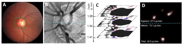
Methods: A 70 kHz spectral-domain OCT system scanned a 2×2 mm area centred at the optic disc of the eyes with DR and healthy participants. The multiplane en face Doppler OCT algorithm generated a three-dimensional volumetric data set consisting of 195 en face planes. The TRBF was calculated from the maximum flow values of each branching retinal vein at an optimised en face plane. DR severity was graded according to the international clinical classification system. The generalised linear model method was used to compare flow values between DR groups and the control group.
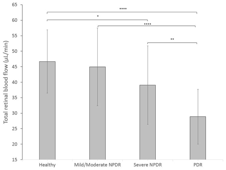
Results: A total of 71 eyes from 71 participants were included. Ten eyes were excluded due to poor image quality. The within-visit repeatability of scans was 4.1% (coefficient of variation). There was no significant difference in the TRBF between the healthy (46.7±10.2 µL/min) and mild/moderate non-proliferative DR (44.9±12.6 µL/min) groups. The TRBF in severe non-proliferative DR (39.1±12.6 µL/min) and proliferative DR (28.9±8.85 µL/min) groups were significantly lower (p=0.04 and p<0.0001, respectively) than that of the healthy group. TRBF was correlated with DR disease severity (p<0.0001, linear trend test).
Conclusion: The novel multiplane en face Doppler OCT method provided reliable measurements of TRBF in DR eyes. This may be a useful tool in understanding the pathophysiology of DR.
Keywords: Diabetic retinopathy; imaging; optical coherence tomography; retina; retinal blood flow.
© Article author(s) (or their employer(s) unless otherwise stated in the text of the article) 2018. All rights reserved. No commercial use is permitted unless otherwise expressly granted.
Conflict of interest statement
Competing interests: Oregon Health & Science University (OHSU), OT, DH and YJ have a significant financial interest in Optovue, a company that may have a commercial interest in the results of this research and technology. OT and DH have a significant financial interest in Carl Zeiss Meditec. These potential conflicts of interest have been reviewed and managed by OHSU.
Figures
Similar articles
-
En Face Doppler Optical Coherence Tomography Measurement of Total Retinal Blood Flow in Diabetic Retinopathy and Diabetic Macular Edema.JAMA Ophthalmol. 2017 Mar 1;135(3):244-251. doi: 10.1001/jamaophthalmol.2016.5774. JAMA Ophthalmol. 2017. PMID: 28196198 Free PMC article.
-
ASSESSMENT OF RETINAL BLOOD FLOW IN DIABETIC RETINOPATHY USING DOPPLER FOURIER-DOMAIN OPTICAL COHERENCE TOMOGRAPHY.Retina. 2017 Nov;37(11):2001-2007. doi: 10.1097/IAE.0000000000001479. Retina. 2017. PMID: 28098726 Free PMC article.
-
Retinal Blood Flow Response to Hyperoxia Measured With En Face Doppler Optical Coherence Tomography.Invest Ophthalmol Vis Sci. 2016 Jul 1;57(9):OCT141-5. doi: 10.1167/iovs.15-18917. Invest Ophthalmol Vis Sci. 2016. PMID: 27409465 Free PMC article.
-
ZEISS Angioplex™ Spectral Domain Optical Coherence Tomography Angiography: Technical Aspects.Dev Ophthalmol. 2016;56:18-29. doi: 10.1159/000442773. Epub 2016 Mar 15. Dev Ophthalmol. 2016. PMID: 27023249 Review.
-
OCT angiography and visible-light OCT in diabetic retinopathy.Vision Res. 2017 Oct;139:191-203. doi: 10.1016/j.visres.2017.05.006. Epub 2017 Jun 21. Vision Res. 2017. PMID: 28601429 Free PMC article. Review.
Cited by
-
Functional OCT angiography reveals early retinal neurovascular dysfunction in diabetes with capillary resolution.Biomed Opt Express. 2023 Mar 27;14(4):1670-1684. doi: 10.1364/BOE.485940. eCollection 2023 Apr 1. Biomed Opt Express. 2023. PMID: 37078055 Free PMC article.
-
Constriction of Retinal Venules to Endothelin-1: Obligatory Roles of ETA Receptors, Extracellular Calcium Entry, and Rho Kinase.Invest Ophthalmol Vis Sci. 2018 Oct 1;59(12):5167-5175. doi: 10.1167/iovs.18-25369. Invest Ophthalmol Vis Sci. 2018. PMID: 30372743 Free PMC article.
-
Decreased ocular blood flow after photocoagulation therapy in neonatal retinopathy of prematurity.Jpn J Ophthalmol. 2017 Nov;61(6):484-493. doi: 10.1007/s10384-017-0536-7. Epub 2017 Sep 20. Jpn J Ophthalmol. 2017. PMID: 28932922
-
Maximum value projection produces better en face OCT angiograms than mean value projection.Biomed Opt Express. 2018 Nov 26;9(12):6412-6424. doi: 10.1364/BOE.9.006412. eCollection 2018 Dec 1. Biomed Opt Express. 2018. PMID: 31065439 Free PMC article.
-
Dorzolamide/Timolol Fixed Combination: Learning from the Past and Looking Toward the Future.Adv Ther. 2021 Jan;38(1):24-51. doi: 10.1007/s12325-020-01525-5. Epub 2020 Oct 27. Adv Ther. 2021. PMID: 33108623 Free PMC article. Review.
References
-
- Curtis TM, Gardiner TA, Stitt AW. Microvascular lesions of diabetic retinopathy: clues towards understanding pathogenesis? Eye. 2009;23:1496–508. - PubMed
-
- Antonetti DA, Barber AJ, Bronson SK, et al. Diabetic retinopathy: seeing beyond glucose-induced microvascular disease. Diabetes. 2006;55:2401–11. - PubMed
-
- Nagaoka T, Sato E, Takahashi A, et al. Impaired retinal circulation in patients with type diabetes mellitus: retinal laser Doppler velocimetry study. Invest Ophthalmol Vis Sci. 2010;51:6729–34. - PubMed
Publication types
MeSH terms
Grants and funding
LinkOut - more resources
Full Text Sources
Other Literature Sources
Medical
Research Materials