

Role of High-Resolution Dynamic Contrast-Enhanced MRI with Golden-Angle Radial Sparse Parallel Reconstruction to Identify the Normal Pituitary Gland in Patients with Macroadenomas
- PMID: 28495945
- PMCID: PMC6080601
- DOI: 10.3174/ajnr.A5244
Role of High-Resolution Dynamic Contrast-Enhanced MRI with Golden-Angle Radial Sparse Parallel Reconstruction to Identify the Normal Pituitary Gland in Patients with Macroadenomas
Abstract
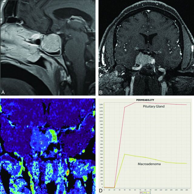
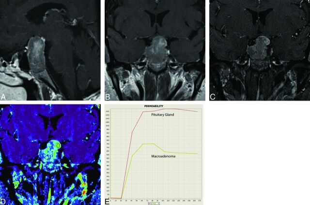
Background and purpose: Preoperative localization of the pituitary gland with imaging in patients with macroadenomas has been inadequately explored. The pituitary gland enhancing more avidly than a macroadenoma has been described in the literature. Taking advantage of this differential enhancement pattern, our aim was to evaluate the role of high-resolution dynamic MR imaging with golden-angle radial sparse parallel reconstruction in localizing the pituitary gland in patients undergoing trans-sphenoidal resection of a macroadenoma.
Materials and methods: A retrospective study was performed in 17 patients who underwent trans-sphenoidal surgery for pituitary macroadenoma. Radial volumetric interpolated brain examination sequences with golden-angle radial sparse parallel technique were obtained. Using an ROI-based method to obtain signal-time curves and permeability measures, 3 separate readers identified the normal pituitary gland distinct from the macroadenoma. The readers' localizations were then compared with the intraoperative location of the gland. Statistical analyses were performed to assess the interobserver agreement and correlation with operative findings.
Results: The normal pituitary gland was found to have steeper enhancement-time curves as well as higher peak enhancement values compared with the macroadenoma (P < .001). Interobserver agreement was almost perfect in all 3 planes (κ = 0.89). In the 14 cases in which the gland was clearly identified intraoperatively, the correlation between the readers' localization and the true location derived from surgery was also nearly perfect (κ = 0.95).
Conclusions: This study confirms our ability to consistently and accurately identify the normal pituitary gland in patients with macroadenomas with the golden-angle radial sparse parallel technique with quantitative permeability measurements and enhancement-time curves.
© 2017 by American Journal of Neuroradiology.
Figures
Similar articles
-
Quantitative pharmacokinetic analysis of high-temporal-resolution dynamic contrast-enhanced MRI to differentiate the normal-appearing pituitary gland from pituitary macroadenoma.Jpn J Radiol. 2020 Jul;38(7):649-657. doi: 10.1007/s11604-020-00942-4. Epub 2020 Mar 11. Jpn J Radiol. 2020. PMID: 32162178
-
High-Resolution DCE-MRI of the Pituitary Gland Using Radial k-Space Acquisition with Compressed Sensing Reconstruction.AJNR Am J Neuroradiol. 2015 Aug;36(8):1444-9. doi: 10.3174/ajnr.A4324. Epub 2015 May 7. AJNR Am J Neuroradiol. 2015. PMID: 25953760 Free PMC article.
-
Golden-angle radial sparse parallel (GRASP) MRI in clinical routine detection of pituitary microadenomas: First experience and feasibility.Magn Reson Imaging. 2019 Jul;60:38-43. doi: 10.1016/j.mri.2019.03.015. Epub 2019 Mar 27. Magn Reson Imaging. 2019. PMID: 30928387
-
"The Pituitary within GRASP" - Golden-Angle Radial Sparse Parallel Dynamic MRI Technique and Applications to the Pituitary Gland.Semin Ultrasound CT MR. 2021 Jun;42(3):307-315. doi: 10.1053/j.sult.2021.04.007. Epub 2021 Apr 22. Semin Ultrasound CT MR. 2021. PMID: 34147165 Review.
-
Pituitary magnetic resonance imaging in Cushing's disease.Endocrine. 2017 Mar;55(3):691-696. doi: 10.1007/s12020-016-1038-y. Epub 2016 Jul 19. Endocrine. 2017. PMID: 27435590 Review.
Cited by
-
Differentiation of Jugular Foramen Paragangliomas versus Schwannomas Using Golden-Angle Radial Sparse Parallel Dynamic Contrast-Enhanced MRI.AJNR Am J Neuroradiol. 2021 Oct;42(10):1847-1852. doi: 10.3174/ajnr.A7243. Epub 2021 Sep 9. AJNR Am J Neuroradiol. 2021. PMID: 34503944 Free PMC article.
-
Quantitative pharmacokinetic analysis of high-temporal-resolution dynamic contrast-enhanced MRI to differentiate the normal-appearing pituitary gland from pituitary macroadenoma.Jpn J Radiol. 2020 Jul;38(7):649-657. doi: 10.1007/s11604-020-00942-4. Epub 2020 Mar 11. Jpn J Radiol. 2020. PMID: 32162178
-
Dynamic Contrast-Enhanced MRI to Differentiate Parotid Neoplasms Using Golden-Angle Radial Sparse Parallel Imaging.AJNR Am J Neuroradiol. 2019 Jun;40(6):1029-1036. doi: 10.3174/ajnr.A6055. Epub 2019 May 2. AJNR Am J Neuroradiol. 2019. PMID: 31048300 Free PMC article.
-
[68 Ga]-DOTATATE PET/MR-based evaluation of physiologic somatostatin receptor 2 expression in the adult pituitary gland as a function of age and sex in a prospective cohort.Pituitary. 2023 Aug;26(4):419-428. doi: 10.1007/s11102-023-01329-0. Epub 2023 Jun 7. Pituitary. 2023. PMID: 37285059
-
MRI Protocol for Pituitary Assessment in Children with Growth or Puberty Disorders-Is Gadolinium Contrast Administration Actually Needed?J Clin Med. 2021 Oct 6;10(19):4598. doi: 10.3390/jcm10194598. J Clin Med. 2021. PMID: 34640616 Free PMC article.
References
MeSH terms
Substances
Grants and funding
LinkOut - more resources
Full Text Sources
Other Literature Sources
Medical