

Neuroimaging Changes in Menkes Disease, Part 1
- PMID: 28495946
- PMCID: PMC7963639
- DOI: 10.3174/ajnr.A5186
Neuroimaging Changes in Menkes Disease, Part 1
Abstract
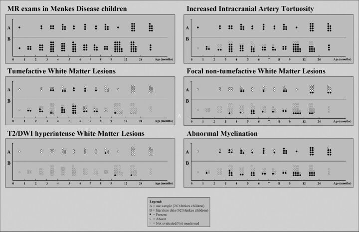
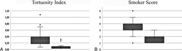
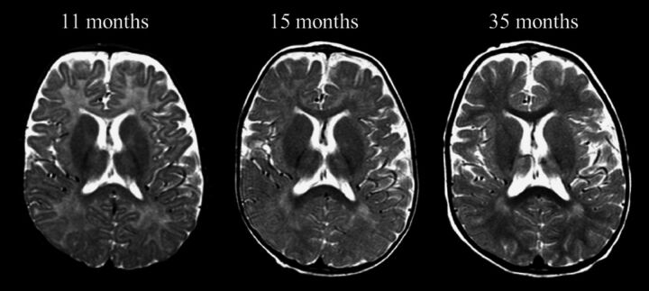
Menkes disease is a rare multisystem X-linked disorder of copper metabolism. Despite an early, severe, and progressive neurologic involvement, our knowledge of brain involvement remains unsatisfactory. The first part of this retrospective and review MR imaging study aims to define the frequency rate, timing, imaging features, and evolution of intracranial vascular and white matter changes. According to our analysis, striking but also poorly evolutive vascular abnormalities characterize the very early phases of disease. After the first months, myelination delay becomes evident, often in association with protean focal white matter lesions, some of which reveal an age-specific brain vulnerability. In later phases of the disease, concomitant progressive neurodegeneration might hinder the myelination progression. The currently enriched knowledge of neuroradiologic finding evolution provides valuable clues for early diagnosis, identifies possible MR imaging biomarkers of new treatment efficacy, and improves our comprehension of possible mechanisms of brain injury in Menkes disease.
© 2017 by American Journal of Neuroradiology.
Figures
References
-
- Van der Knaap MS, Valk J. Magnetic Resonance of Myelination and Myelin Disorders Disorders. Berlin: Springer; 2005
Publication types
MeSH terms
LinkOut - more resources
Full Text Sources
Other Literature Sources