. 2017 Jul;206(3):1459-1468.
doi: 10.1534/genetics.117.200576. Epub 2017 May 11.

Maternal Torso-Like Coordinates Tissue Folding During Drosophila Gastrulation

Affiliations

Maternal Torso-Like Coordinates Tissue Folding During Drosophila Gastrulation

Travis K Johnson et al. Genetics. 2017 Jul.

Abstract

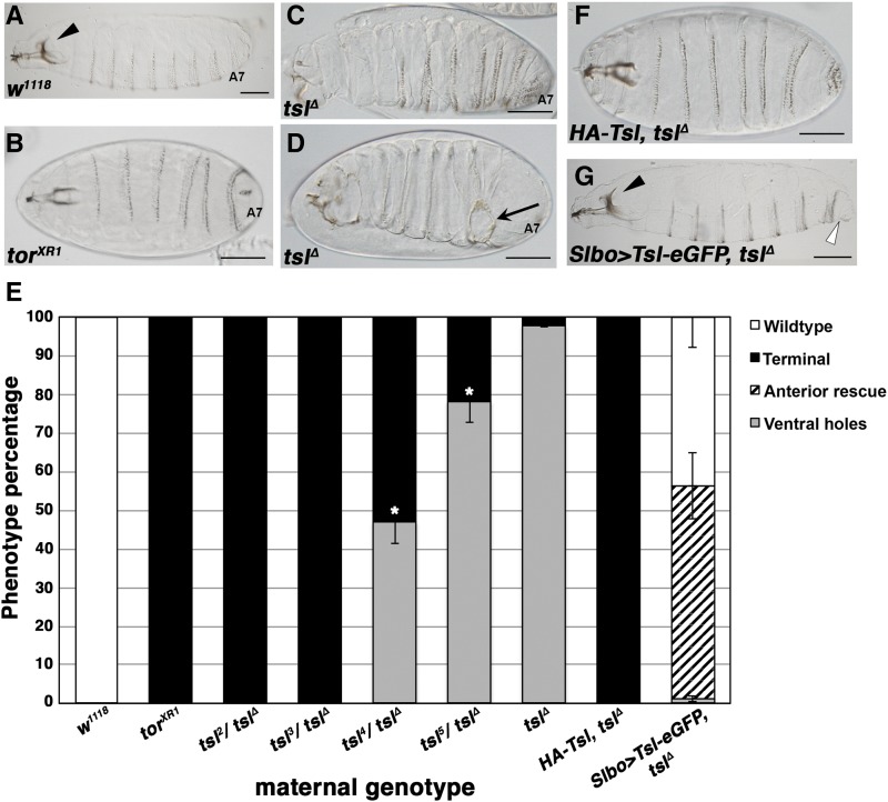
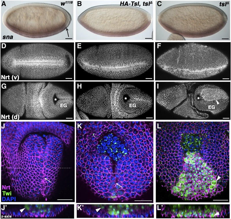
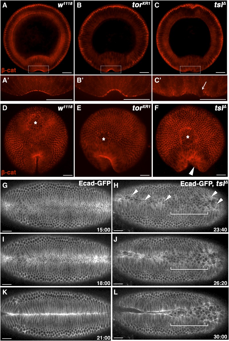
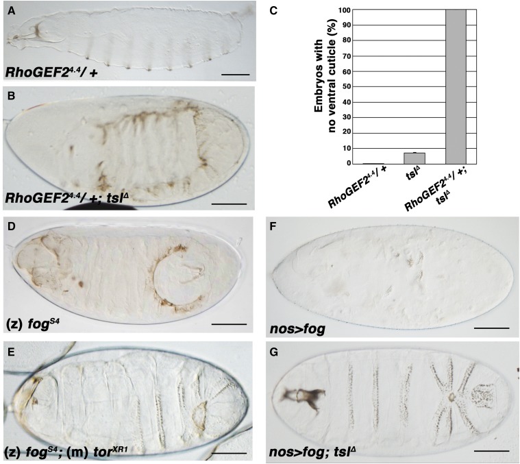
The rapid and orderly folding of epithelial tissue during developmental processes such as gastrulation requires the precise coordination of changes in cell shape. Here, we report that the perforin-like protein Torso-like (Tsl), the key extracellular determinant for Drosophila embryonic terminal patterning, also functions to control epithelial morphogenesis. We find that tsl null mutants display a ventral cuticular hole phenotype that is independent of the loss of terminal structures, and arises as a consequence of mesoderm invagination defects. We show that the holes are caused by uncoordinated constriction of ventral cell apices, resulting in the formation of an incomplete ventral furrow. Consistent with these data, we find that loss of tsl is sensitive to gene dosage of RhoGEF2, a critical mediator of Rho1-dependent ventral cell shape changes during furrow formation, suggesting that Tsl may act in this pathway. In addition, loss of tsl strongly suppressed the effects of ectopic expression of Folded Gastrulation (Fog), a secreted protein that promotes apical constriction. Taken together, our data suggest that Tsl controls Rho1-mediated apical constriction via Fog. Therefore, we propose that Tsl regulates extracellular Fog activity to synchronize cell shape changes and coordinate ventral morphogenesis in Drosophila Identifying the Tsl-mediated event that is common to both terminal patterning and morphogenesis will be valuable for our understanding of the extracellular control of developmental signaling by perforin-like proteins.

Keywords: Drosophila; MACPF; Torso-like; gastrulation; morphogenesis.


Figures

Figure 1
Figure 1
Loss of maternal tsl results in ventral cuticular holes that are independent of terminal patterning failure. (A) Wild-type larval cuticle with complete head skeleton (arrowhead) and abdominal segment 7 (A7) present. (B) Cuticle of embryos laid by torXR1 females showing the terminal class mutant phenotype. Note that the head skeleton is reduced and structures posterior to A7 are absent. (C and D) Cuticles of embryos laid by tslΔ mothers display the terminal phenotype (C) similar to torXR1; however, a large proportion also possess ventral cuticular holes (D, arrow). (E) Quantification of phenotypes observed in embryos laid by mothers of the genotypes shown. Hypomorphic alleles of tsl in trans with the null allele (tslΔ) form an allelic series with respect to the cuticular hole phenotype. Asterisks indicate significant differences from the tslΔ phenotype (t-test, P < 0.05). Means are plotted and error bars represent one SE calculated from at least three cuticle preparations (> 100 cuticles scored for each). (F) A genomic transgene containing HA-tagged tsl (HA-Tsl) completely rescues the cuticular hole phenotype of tslΔ; however, it does not restore terminal patterning. (G) Expression of an enhanced GFP (eGFP)-tagged tsl transgene in the endogenous ovarian pattern of tsl (Slbo-Gal4) restores the tslΔ ventral cuticle and partially restores terminal patterning, as assessed by rescue of the anterior head skeleton and presence of abdominal segment 8. The cuticle shown is a representative image where the addition of A8 tissue (open arrowhead) and a wild-type head skeleton (arrowhead) is observed. Anteriors are to the left. Bar, 100 μm.
Figure 2
Figure 2
The ventral cuticular holes in tslΔ embryos are caused by impaired mesoderm invagination during morphogenesis. Wild-type (w1118, left panels), terminal class mutant (HA-Tsl, tslΔ, center panels), and tslΔ (right panels) embryos stained with various markers of embryogenesis. (A–C) Transcript expression of the ventral cell fate marker Snail (sna) is repressed at the posterior of wild-type embryos by the terminal system (A, arrow). In tslΔ (B) and terminal class mutant (C) embryos, sna expression extends to the posterior pole. (D–F) Ventral (v) furrow formation in gastrulating embryos labeled with the membrane marker anti-Neurotactin (Nrt). Wild-type (D) and terminal class mutant (E) embryos form regular ventral furrows. The furrows from tslΔ embryos are irregular and incomplete (F). (G–I) Dorsal (d) views of gastrulated embryos stained with anti-Nrt. (G) Wild-type embryos correctly invaginate their posterior midgut (pole cell position indicated by asterisks) unlike terminal class mutant (H) and tslΔ (I) embryos due to terminal system failure. however, tslΔ embryos possess a large field of intensely labeled cells on the extended germband (EG) that are not seen in wild-type nor terminal class mutant embryos (arrowhead). Anteriors are to the left. (J–L) Posterior–dorsal surface views of gastrulae embryos stained with anti-Nrt (magenta), anti-Twist (Twi, green) to label mesodermal precursors, and DAPI (blue). The ventral furrow has closed at the midline (white arrow) in wild-type (J) terminal class mutant (K) embryos but remains open in tslΔ embryos (L), as indicated by the surface location of Twi-positive cells (white arrowheads). Anteriors are to the top. Lower panels (J’–L’) show the z-axis cross-section of the top panels at the position indicated by the dotted white line. Twi-positive nuclei are visible only below the dorsal surface in wild-type (J’) and terminal class mutant (K’) embryos indicating successful furrow invagination. Many of these cells remain at the dorsal surface of tslΔ embryos (L’). Maternal genotypes are shown. Bar, 50 μm.
Figure 3
Figure 3
Tsl promotes and coordinates apical constriction during ventral furrow formation. (A–C) Cross-sections of fixed early gastrulae embryos sectioned at approximately two-thirds the embryo length (from the anterior to posterior) and labeled with anti-β-catenin to reveal adherens junctions (AJs). β-catenin localizes strongly to the apical surface in the early ventral furrow in wild-type (w1118, A) and terminal class mutant (torXR1, B) embryos, but poorly and irregularly in tslΔ embryos (C). Lower panels are (A’–C’) high-magnification images of the boxed area in the top panels. Areas of low apical β-catenin correspond to unconstricted cells indicated by apical nuclei (arrowed). Posterior view of embryos with closing ventral furrows. Uniform furrows and concentrated apical AJs are observed for wild-type (D) and terminal class mutant (E) embryos, despite the latter failing to internalize the posterior midgut (asterisks) and forming a posteriorly extended furrow as expected. tslΔ embryos fail similarly in these regards; however, they also display an irregular shaped posterior furrow (F, arrowhead). Ventral is to the bottom. (G–L) Live imaging stills taken at 3-min intervals of representative Ecad-GFP-expressing control (G, I, and K) and tslΔ (H, J, and L) embryos during ventral furrow formation and invagination. Furrow formation in tslΔ embryos is delayed by ∼8 min compared to controls. Cell constrictions in the ventral domain of tslΔ embryos are limited to sporadic patches of cells (arrowheads). Areas containing more constricted cells appear to initiate furrow formation first. Invagination is incomplete in this example due to a large population of cells toward the posterior (bracketed) remaining unconstricted. Time is in minutes postcellularization completion. Ventral side is shown with anteriors to the left. Maternal genotypes are indicated. Bar, 35 μm.
Figure 4
Figure 4
tsl interacts with RhoGEF2 and fog in ventral morphogenesis. (A) Progeny from RhoGEF24.4 heterozygote females display a wild-type cuticle pattern. (B) Progeny from females heterozygous for RhoGEF24.4 and lacking tsl (tslΔ) display a severe loss of ventral cuticle. (C) A striking increase in the proportion of embryos lacking ventral cuticles is observed when RhoGEF2 dosage is reduced in a tslΔ background compared to tslΔ alone (tslΔ = 7.3% vs. RhoGEF24.4/+; tslΔ = 100%). Means are plotted and error bars represent one SE calculated from at least three cuticle preparations (> 100 cuticles scored for each). (D) Posterior–ventral holes are observed in cuticles of fogs4 embryos. (E) fogs4;torXR1 double-mutant embryos have a cuticle phenotype that closely resembles that of tslΔ. (F) Embryos from females expressing fog ubiquitously from the maternal germline (nos>) are severely compromised in their ability to produce ventral cuticle. (G) The cuticle phenotype of embryos that express fog in the absence of tsl closely resembles the tslΔ phenotype. Maternal genotypes are shown unless otherwise indicated (m, maternal; z, zygotic). Anteriors are to the left, ventral is down. Bar, 100 μm.

Similar articles

Cited by

References

    1. Barrett K., Leptin M., Settleman J., 1997. The Rho GTPase and a putative RhoGEF mediate a signaling pathway for the cell shape changes in Drosophila gastrulation. Cell 91: 905–915. - PubMed
    1. Bischof J., Maeda R. K., Hediger M., Karch F., Basler K., 2007. An optimized transgenesis system for Drosophila using germ-line-specific phiC31 integrases. Proc. Natl. Acad. Sci. USA 104: 3312–3317. - PMC - PubMed
    1. Casanova J., Struhl G., 1989. Localized surface activity of torso, a receptor tyrosine kinase, specifies terminal body pattern in Drosophila. Genes Dev. 3: 2025–2038. - PubMed
    1. Costa M., Wilson E. T., Wieschaus E., 1994. A putative cell signal encoded by the folded gastrulation gene coordinates cell shape changes during Drosophila gastrulation. Cell 76: 1075–1089. - PubMed
    1. Cox R. T., Kirkpatrick C., Peifer M., 1996. Armadillo is required for adherens junction assembly, cell polarity, and morphogenesis during Drosophila embryogenesis. J. Cell Biol. 134: 133–148. - PMC - PubMed