. 2017 Jun 7;37(23):5670-5680.
doi: 10.1523/JNEUROSCI.2968-16.2017. Epub 2017 May 11.

Optogenetic Inhibition of Ventral Pallidum Neurons Impairs Context-Driven Salt Seeking

Affiliations

Optogenetic Inhibition of Ventral Pallidum Neurons Impairs Context-Driven Salt Seeking

Stephen E Chang et al. J Neurosci. .

Abstract

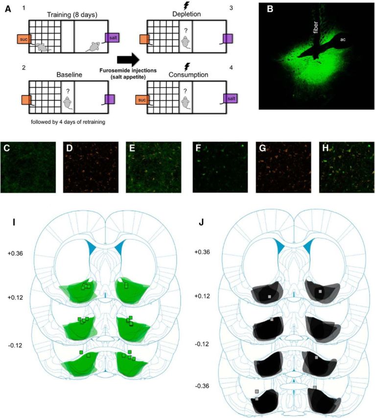
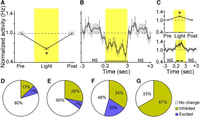
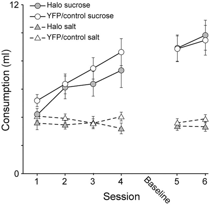
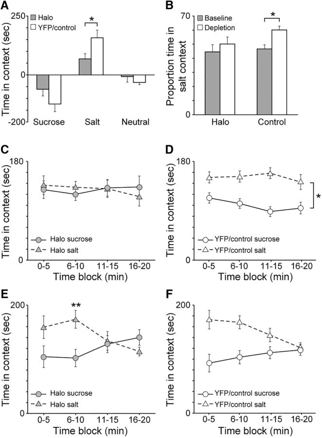
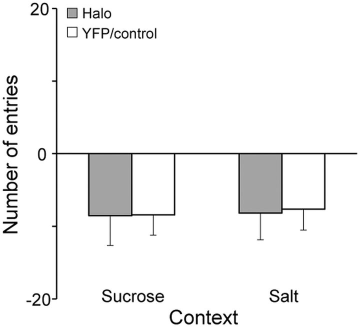
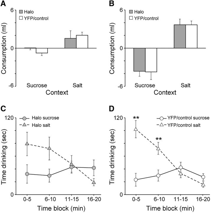
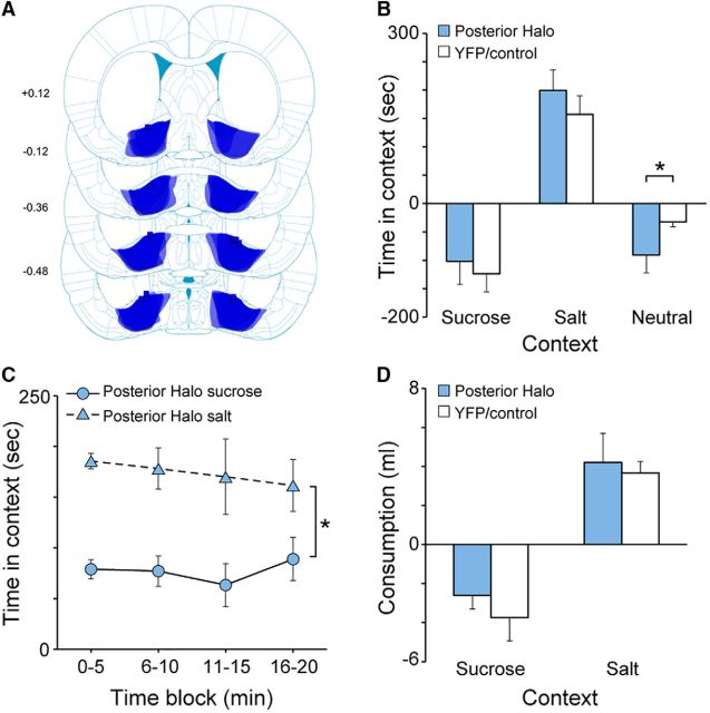
Salt appetite, in which animals can immediately seek out salt when under a novel state of sodium deprivation, is a classic example of how homeostatic systems interface with learned associations to produce an on-the-fly updating of motivated behavior. Neural activity in the ventral pallidum (VP) has been shown to encode changes in the value of salt under such conditions, both the value of salt itself (Tindell et al., 2006) and the motivational value of its predictive cues (Tindell et al., 2009; Robinson and Berridge, 2013). However, it is not known whether the VP is necessary for salt appetite in terms of seeking out salt or consuming salt following sodium depletion. Here, we used a conditioned place-preference procedure to investigate the effects of optogenetically inhibiting the VP on context-driven salt seeking and the consumption of salt following deprivation. Male rats learned to associate one context with sucrose and another context with less-desirable salt. Following sodium depletion, and in the absence of either sucrose or salt, we found that inhibiting the VP selectively reduced the elevation in time spent in the salt-paired context. VP inhibition had minimal effects on the consumption of salt once it was made available. To our knowledge, this is the first evidence that the VP or any brain region is necessary for the ability to use contextual cues to guide salt seeking. These results highlight a dissociation between deficit-driven reward seeking and reward consumption to replenish those deficits, with the former process being particularly sensitive to on-line VP activity.SIGNIFICANCE STATEMENT Salt appetite, in which rats will immediately seek out a once-undesirable concentrated salt solution after being depleted of bodily sodium despite never having tasted salt as a positive reward, is a phenomenon showing how animals can update their motivational goals without any new learning or conditioning. This salt-seeking behavior is also observed when the animal is presented with salt-paired cues. The neural circuitry necessary for context-driven salt-seeking behavior is unknown. We used a novel conditioned place preference procedure to show that optogenetic inhibition of the ventral pallidum (VP), a region known for processing reward, impairs context-driven salt seeking and has minimal effects on the consumption of salt itself following sodium depletion. These results highlight the importance of the VP in context-driven reward-seeking behavior.

Keywords: salt appetite; ventral pallidum.


Figures

Figure 1.
Figure 1.
A, Behavioral procedure: rats were trained to associate one context with access to sucrose and another context with access to salt (4 d in each context, 20 min per session). Rats were then given a baseline test in which they were allowed access to both contexts for the first time in the absence of sucrose or salt (flavored solutions still present). After 4 d of retraining (2 d in each context), rats were sodium-depleted with furosemide (10 mg/kg, i.p.). After 48 h, rats were given a depletion test identical to the baseline test except that now rats received yellow laser stimulation (3 s on, 3 s off train). Finally, rats were given a consumption test identical to the depletion test but now sucrose and salt were available in each solution. B, Representative brain slice from a Halo rat showing eNpHR3.0-EYFP expression in the VP and fiber track. ac, Anterior commissure. C–E, Example of eNpHR3.0-EYFP, Neurotrace, and coexpression of each from a brain slice of a rat that received AAV5-hSyn-eNpHR3.0-EYFP infusions in the VP. F–H, Example of EYFP, Neurotrace, and coexpression of each from a brain slice of a rat that received AAV5-hSyn-EYFP infusions in the VP. I, Schematic diagram of eNpHR3.0-EYFP expression in the VP (shading overlay) and fiber implant placements (squares) from all Halo rats (n = 12). Numbers on the left are number of millimeters from bregma. Illustrations adapted from Paxinos and Watson (2009). J, Schematic diagram of eNpHR3.0-EYFP expression in the VP (shading overlay) and fiber implant placements (squares) from all rats classified as Misses (n = 5). Note that three placements are not mapped due to either the fiber placement being too anterior and not above or within the VP (beyond 0.36 mm from bregma; n = 2) or the fiber placement was unable to be located (n = 1). Numbers on the left are number of millimeters from bregma. Illustrations adapted from Paxinos and Watson (2009).
Figure 2.
Figure 2.
A, Normalized activity of inhibited cells (n = 12) during the 3 s period before light delivery (Pre-Light), during light delivery (Light), and during the 3 s period after light delivery (Post-Light). Note the data for the Pre-Light and Post-Light periods are identical due to the continuous light on/off delivery cycle (3 s on/off). B, Activity of inhibited cells during the Pre-Light, Light, and Post-Light periods in 200 ms bins. C, Top, Normalized activity of excited cells (n = 7) during the Pre-Light, Light, and Post-Light delivery periods. Bottom, Activity of excited cells during the Pre-Light, Light, and Post-Light periods in 200 ms bins. D, Proportion of inhibited units out of total recorded units. E, Proportion of inhibited units out of total recorded units on tetrodes that ever had an inhibited unit on any recording session. F, Proportion of inhibited units out of total recorded units from tetrodes that had an inhibited or excited unit on that same recording session. G, Proportion of inhibited units out of total recorded units from the tetrodes on which the inhibited unit was recorded during each session. Error bars represent the SEM. *p < 0.05 compared with baseline average. ns, Not significant.
Figure 3.
Figure 3.
Amount of sucrose and salt solutions consumed over the first 4 d (1–4) and the last 2 d (5, 6) of training following the baseline test session (Baseline). There were no group differences between Halo and YFP/Control rats. Error bars represent ±SEM.
Figure 4.
Figure 4.
A, Elevation in time spent in sucrose-paired and salt-paired contexts during the depletion test following sodium depletion compared with the baseline test. Compared with Halo rats, YFP/Control rats showed a greater elevation in time spent in the salt-paired context. There was no difference in time spent in the sucrose-paired context. B, The proportion of time spent in the salt-paired context relative to the time spent in both the sucrose-paired and salt-paired contexts during the baseline and depletion tests. YFP/Control rats spent a greater proportion of time in the salt-paired context following sodium depletion during the depletion test compared with the baseline test. Halo rats showed no differences. C, Amount of time Halo rats spent in the sucrose-paired and salt-paired contexts during the depletion test in 5 min blocks. Halo rats did not show any differences in time spent in one context versus the other context across the entire session. D, Amount of time YFP/Control rats spent in the sucrose-paired and salt-paired contexts during the depletion test in 5 min blocks. Control rats spent more time in the salt-paired context compared with the sucrose-paired context across the entire session. E, Amount of time Halo rats spent in the sucrose-paired and salt-paired contexts during the consumption test in 5 min blocks. Halo rats spent significantly more time in the salt-paired context than in the sucrose-paired context during only the second block. F, Amount of time YFP/Control rats spent in the sucrose-paired and salt-paired contexts during the consumption test in 5 min blocks. YFP/Control rats spent significantly more time in the salt-paired context than in the sucrose-paired context during the first two blocks. Error bars represent the SEM. *p < 0.05; **p < 0.0125 after Bonferroni's correction.
Figure 5.
Figure 5.
Elevation in the number of entries into the sucrose-paired and salt-paired contexts during the depletion test following sodium depletion compared with the baseline test. Halo and YFP/Control rats showed no differences. Error bars represent the SEM.
Figure 6.
Figure 6.
A, Elevation in the amount of solution consumed (without sucrose or salt) during the depletion test following sodium depletion compared with the baseline test. Although there were no differences between groups in terms of consumption of each flavor, YFP/Control rats consumed significantly more salt-paired flavor than sucrosepaired flavor. Halo rats also consumed more salt-paired flavor than sucrose-paired flavor, but the difference was not statistically significant. B, Elevation in the amount of solution consumed (with sucrose and salt) during the consumption test following sodium depletion compared with the last day of training. Halo and YFP/Control rats showed no differences. C, Amount of time Halo rats spent consuming sucrose-paired and salt-paired solutions during the consumption test (when sucrose and salt were made available) in 5 min blocks. Halo rats did not show any statistical differences after correction for multiple comparisons. D, Amount of time YFP/Control rats spent consuming sucrose-paired and salt-paired solutions during the consumption test (when sucrose and salt were made available) in 5 min blocks. YFP/Control rats spent significantly more time drinking the salt-paired solution compared with the sucrose-paired solution during the first two blocks. Error bars represent the SEM. **p < 0.0125 after Bonferroni's correction.
Figure 7.
Figure 7.
A, Schematic diagram of eNpHR3.0-EYFP expression in the VP (shading overlay) and fiber implant placements (squares) from all Posterior Halo rats (n = 5). Numbers on the left are number of millimeters from bregma. Illustrations adapted from Paxinos and Watson (2009). B, Elevation in time spent in sucrose-paired and salt-paired contexts during the depletion test following sodium depletion compared with the baseline test. C, Amount of time Posterior Halo rats spent in the sucrose-paired and salt-paired contexts during the depletion test in 5 min blocks. Posterior Halo rats spent more time in the salt-paired context than in the sucrose-paired context throughout the entire test session. D, Elevation in the amount of solutions consumed (with sucrose or salt) during the consumption test following sodium depletion compared with the last day of training. Error bars represent the SEM.

References

    1. Ahrens AM, Meyer PJ, Ferguson LM, Robinson TE, Aldridge JW (2016) Neural activity in the ventral pallidum encodes variation in the incentive value of a reward cue. J Neurosci 36:7957–7970. 10.1523/jneurosci.0736-16.2016 - DOI - PMC - PubMed
    1. Anikeeva P, Andalman AS, Witten I, Warden M, Goshen I, Grosenick L, Gunaydin LA, Frank LM, Deisseroth K (2011) Optetrode: a multichannel readout for optogenetic control in freely moving mice. Nat Neurosci 15:163–170. 10.1038/nn.2992 - DOI - PMC - PubMed
    1. Chang SE, Todd TP, Bucci DJ, Smith KS (2015) Chemogenetic manipulation of ventral pallidal neurons impairs acquisition of sign-tracking in rats. Eur J Neurosci 42:3105–3116. 10.1111/ejn.13103 - DOI - PMC - PubMed
    1. Cromwell HC, Berridge KC (1993) Where does damage lead to enhanced food aversion: the ventral pallidum/substantia innominata or lateral hypothalamus? Brain Res 624:1–10. 10.1016/0006-8993(93)90053-P - DOI - PubMed
    1. Dallimore JE, Mickiewicz AL, Napier TC (2006) Intra-ventral pallidal glutamate antagonists block expression of morphine-induced place preference. Behav Neurosci 120:1103–1114. 10.1037/0735-7044.120.5.1103 - DOI - PubMed

Publication types

Substances