. 2017 May 11;7(1):1744.
doi: 10.1038/s41598-017-01591-w.

'Gardos Channelopathy': a variant of hereditary Stomatocytosis with complex molecular regulation

Affiliations

'Gardos Channelopathy': a variant of hereditary Stomatocytosis with complex molecular regulation

Elisa Fermo et al. Sci Rep. .

Abstract

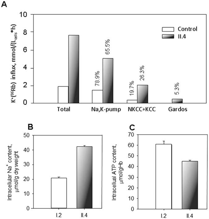
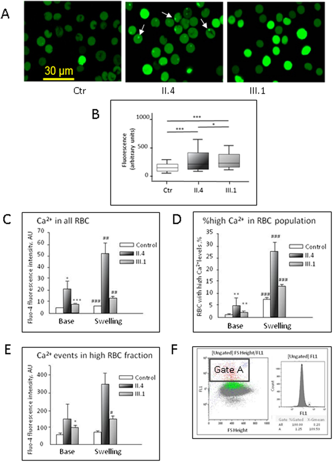
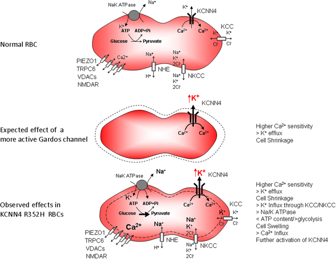
The Gardos channel is a Ca2+ sensitive, K+ selective channel present in several tissues including RBCs, where it is involved in cell volume regulation. Recently, mutations at two different aminoacid residues in KCNN4 have been reported in patients with hereditary xerocytosis. We identified by whole exome sequencing a new family with two members affected by chronic hemolytic anemia carrying mutation R352H in the KCNN4 gene. No additional mutations in genes encoding for RBCs cytoskeletal, membrane or channel proteins were detected. We performed functional studies on patients' RBCs to evaluate the effects of R352H mutation on the cellular properties and eventually on the clinical phenotype. Gardos channel hyperactivation was demonstrated in circulating erythrocytes and erythroblasts differentiated ex-vivo from peripheral CD34+ cells. Pathological alterations in the function of multiple ion transport systems were observed, suggesting the presence of compensatory effects ultimately preventing cellular dehydration in patient's RBCs; moreover, flow cytometry and confocal fluorescence live-cell imaging showed Ca2+ overload in the RBCs of both patients and hypersensitivity of Ca2+ uptake by RBCs to swelling. Altogether these findings suggest that the 'Gardos channelopathy' is a complex pathology, to some extent different from the common hereditary xerocytosis.


Conflict of interest statement

The authors declare that they have no competing interests.

Figures

Figure 1
Figure 1
(A) LoRRca Osmoscan profile (grey area refers to 150 normal controls); (B) Filterabilty results; healthy cell untreated (Ctr, white bar), pre- incubated with GsMTx-4 (upward hatched bar), TRAM-34 (downward hatched bar) GsMTx-4 + TRAM-34 (dotted bar); patient II.4 (black shaded bar); patient III.1 (grey bar). All measurement were performed at least twice and significance was testes using an unpaired t-test. ** and ***denote p < 0.01 and 0.001 respectively; (C) Separation of RBC on Percoll density gradient and % of cells in low density (L), medium (M) and high (D) density fractions.
Figure 2
Figure 2
(A) Inheritance pattern of the family. (B) Schematic representation of KCNN4 protein domains, and position of R352H mutation.
Figure 3
Figure 3
Whole-cell recordings of KCNN4 (Gardos channel) currents in RBCs from healthy donors and patient II.4. Currents were elicited by voltage steps from −130 mV to 70 mV for 500 ms in 20 mV increments at Vh = −30 mV and recorded in the absence and after application of 1 µM TRAM-34, a specific Gardos channel blocker. (A) Raw current traces from healthy and mutated RBCs in the absence (basal) and in the presence of 1 µM TRAM-34 as indicated above the recordings. For clarity, in all the panels, not all the traces, but every second one, starting with −130 mV, are being shown. (B) Corresponding I/V-curves in the absence of 1 µM TRAM-34, basal (grey diamonds) and in the presence of 1 µM TRAM-34 (black triangles) for healthy (Ctr) and mutated (II.4) RBCs. Data are expressed as mean current ± SEMs. (C) Differences in Gardos currents between healthy (n = 27 cells) and mutated RBCs (n = 23 cells) at −110 mV. (Ca) compares the percent of block (by 1 µM TRAM-34) of mean currents in healthy and mutated RBCs. Due to the small number and small single channel conductivity of Gardos channels in RBCs an additional assessment of Gardos currents is given by analysis of the kinetics of current traces. A mean whole-cell current being the result from the summation of many smaller unit currents flowing through single ion channels, exhibits fluctuations or “noise” about its mean level. (Cb) compares the difference in the standard deviation (SD) of current traces (with and without 1 µM TRAM-34) between control and patient RBCs. Significance was checked based on an unpaired t-test *p < 0.05.
Figure 4
Figure 4
Current-voltage plots for the erythroid precursor cells of the healthy controls (n = 17, black circles) and II-4 patient (open or grey symbols). (A) Cells were exposed to a ramp protocol three times in a row. Each next hyperpolarization-depolarization cycle was followed by an increase in current at positive potentials in the EPCs of II.4 patient (n = 24, 19 and 12 for the ramps 1, 2 and 3), whereas for the cells derived from healthy donors it was never the case. (B) Increase in electric current at positive potential in cells of patient II.4 caused by repetitive ramp protocol application could be blocked by pre-treatment of cells with 15 µM methyl isobutyl amiloride (MIA, grey squares, n = 14), 15 µM TRAM-34 (black squares, n = 7) or omission of the extracellular Ca2+ (open triangles, n = 14). All I/V curves presented in the panel represent the values obtained for the third ramp protocol. Data are shown as mean ± SD.
Figure 5
Figure 5
Activity of ion transporters, intracellular Na+ and ATP content in circulating RBCs. (A) Unidirecitonal K+(86Rb+) influx into RBCs of a healthy control and patient II.4. Shown in the panel are bulk K+ influx and the influx components carried by the Na,K-pump (ouabain-sensitive flux) and the fluxes mediated by the chloride-dependent influx mediated by Na,K,2Cl-, K,Cl-cotransporters, and Gardos channel activity (TRAM-34-sensitive flux). White bars show the flux values for RBCs of healthy control and black bars are for the fluxes in II.4 patient’s RBCs. Numbers above the bars indicate the contribution of each ion transporter into the total influx of either patient or healthy control in %. Intracellular Na+ (B) and ATP (C) content of RBCs of the healthy I.2 (white bar) and patient II.4 (black bar) measured in triplicates on one occasion. Data are mean ± SD. Although statistical analysis could not be performed for single experiments all the values show clear difference between the I.2 and II.4 (for intracellular Na+ and ATP levels) as well between the RBCs of II.4 and identically treated healthy control for all the K+(86Rb+) flux components.
Figure 6
Figure 6
Calcium handling in patients with mutated Gardos channel. (A) Representative confocal images of RBCs from controls and the patients loaded with the Ca2+-sensitive dye Fluo-4. A brighter fluorescence corresponds to a higher Ca2+-concentration. White arrows indicate sequestration of Ca2+ in intracellular vescicles. (B) Statistical analysis of wide field fluorescence Fluo-4 recordings for healthy donors (white bar, n = 2495 cells), patient II.4 (black shaded bar, n = 1257 cells) and patient III.1 (grey bar, n = 1402 cells). Because values are not Gaussian distributed we plotted boxes with whiskers from the 10th to 90th percentile. Significance was checked using the Mann-Whitney test; *, ***denote p < 0.05, and 0.001 respectively. (CF) Intracellular Ca2+ (assessed as Ca2+-dependent fluorescence of Fluo-4) in RBCs of healthy controls and patients II.4 and III.1 measured by flow cytometry. Intracellular Ca2+ was measured at baseline and then the measurements were repeated immediately after the initiation of osmotic swelling by adding a bolus of distilled water to dilute the RBC suspension in isosmotic buffer by 1/3 (Swelling). (C) Readout for all RBCs in suspension; (D) Amount of cells forming “high Ca2+ fraction” (A-gated fraction, flow cytometric analysis shown in panel (E); (F) fluorescence intensity of Fluo-4 in this “high Ca2+ fraction”. All the experiments were performed in two occasions, and each time triple measurements were performed for each conditions. *, ** and ***denote p < 0.05, 0.01 and 0.001 respectively compared to healthy control. #, ## and ### stand for p < 0.05, 0.01 and 0.001 for osmotically compromised cells compared to the baseline values in either control or patients.
Figure 7
Figure 7
Schematic representation of the proposed mechanism observed in RBCs from patient carrying R352H KCNN4 mutation. Top: Normal RBC; Middle: Expected effect of a more active Gardos Channel, Bottom: observed effects in KCNN4 R352H mutant RBCs.

References

    1. Maher AD, Kuchel PW. The Gárdos channel: a review of the Ca2+-activated K+ channel in human erythrocytes. Int J Biochem Cell Biol. 2003;35:1182–1197. doi: 10.1016/S1357-2725(02)00310-2. - DOI - PubMed
    1. Fanger CM, et al. Calmodulin mediates calcium-dependent activation of the intermediate conductance KCa channel, IKCa1. J Biol Chem. 1999;274:5746–5754. doi: 10.1074/jbc.274.9.5746. - DOI - PubMed
    1. Dyrda A, et al. Local membrane deformations activate Ca2+-dependent K+ and anionic currents in intact human red blood cells. PLoS One. 2010;5:e9447. doi: 10.1371/journal.pone.0009447. - DOI - PMC - PubMed
    1. Wulff H, Köhler R. Endothelial small-conductance and intermediate-conductance KCa channels: an update on their pharmacology and usefulness as cardiovascular targets. J Cardiovasc Pharmacol. 2013;61:102–112. doi: 10.1097/FJC.0b013e318279ba20. - DOI - PMC - PubMed
    1. Wulff H, Castle NA. Therapeutic potential of KCa3.1 blockers: recent advances and promising trends. Expert Rev Clin Pharmacol. 2010;3:385–396. doi: 10.1586/ecp.10.11. - DOI - PMC - PubMed

Publication types

MeSH terms

Substances

Supplementary concepts