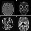
Minimally invasive endoscopic resection for the treatment of sinonasal malignancy: the outcomes and risk factors for recurrence
- PMID: 28496329
- PMCID: PMC5422458
- DOI: 10.2147/TCRM.S131185
Minimally invasive endoscopic resection for the treatment of sinonasal malignancy: the outcomes and risk factors for recurrence
Abstract
Purpose: The role of minimally invasive endoscopic resection (MIER) in the treatment of sinonasal malignancy is controversial. Herein, we performed a retrospective review of a large case series of sinonasal malignancy patients treated with MIER aimed at evaluating the outcomes and identifying the risk factors for recurrence.
Methods: Patients with sinonasal malignancy who underwent MIER from March 2000 to May 2015 were enrolled, and their clinical data were collected. The clinical outcomes were evaluated by determining the 5-year overall survival (OS) and disease-free survival (DFS). The predictive factors for survival and potential independent risk factors for recurrence were explored.
Results: A total of 120 patients were enrolled, including 62 males and 58 females. The mean follow-up period was 51.4 (95% confidence interval: 44.0-59.1) months. The most frequent histological type was mucosal malignant melanoma. The positive margin rate was 19.2% (23/120). Seventy-one patients had the safety anatomic plane (SAP). Age ≥50 years, nodal metastasis, and not having the SAP were found to be predictive factors for survival, and absence of SAP was found to be an independent risk factor for recurrence.
Conclusion: Our study indicated that MIER is an effective and safe surgical procedure in appropriately selected patients. Tumor resection with a safety anatomic boundary is likely to lead to improved survival and decreased recurrence. However, a larger sample and long-term prospective observation are still required to establish the role of MIER in treatment of sinonasal malignancy.
Keywords: malignancy; minimally invasive endoscopic resection; outcome; recurrence; sinonasal skull base.
Conflict of interest statement
Disclosure The authors report no conflicts of interest in this work.
Figures


Similar articles
-
Minimally invasive endoscopic resection of sinonasal undifferentiated carcinoma.Am J Otolaryngol. 2011 Nov-Dec;32(6):464-9. doi: 10.1016/j.amjoto.2010.09.006. Epub 2010 Oct 30. Am J Otolaryngol. 2011. PMID: 21041001
-
Prognostic value of surgical margins during endoscopic resection of paranasal sinus malignancy.Int Forum Allergy Rhinol. 2015 May;5(5):454-9. doi: 10.1002/alr.21463. Epub 2015 Mar 10. Int Forum Allergy Rhinol. 2015. PMID: 25758938
-
Long-term changes in quality of life after endoscopic resection of sinonasal and skull-base tumors.Int Forum Allergy Rhinol. 2015 Dec;5(12):1129-35. doi: 10.1002/alr.21608. Epub 2015 Aug 6. Int Forum Allergy Rhinol. 2015. PMID: 26249825
-
Craniofacial resection of malignant tumors of the anterior skull base: a case series and a systematic review.Acta Neurochir (Wien). 2018 Dec;160(12):2339-2348. doi: 10.1007/s00701-018-3716-4. Epub 2018 Nov 7. Acta Neurochir (Wien). 2018. PMID: 30402666
-
Development of minimally invasive surgery for sinonasal malignancy.Eur Ann Otorhinolaryngol Head Neck Dis. 2016 Dec;133(6):405-411. doi: 10.1016/j.anorl.2016.06.001. Epub 2016 Jul 4. Eur Ann Otorhinolaryngol Head Neck Dis. 2016. PMID: 27386803 Review.
Cited by
-
Endoscopic Craniofacial Resection, an Emerging Epitome of Anterior Skull Base Tumor Resection.Indian J Surg Oncol. 2025 Feb;16(1):70-77. doi: 10.1007/s13193-024-02031-8. Epub 2024 Jul 22. Indian J Surg Oncol. 2025. PMID: 40114904
-
Surgical Interventions for Advanced Parameningeal Rhabdomyosarcoma of Children and Adolescents.Cureus. 2018 Jan 9;10(1):e2045. doi: 10.7759/cureus.2045. Cureus. 2018. PMID: 29541566 Free PMC article. Review.
References
-
- Stammberger H, Anderhuber W, Walch C, Papaefthymiou G. Possibilities and limitations of endoscopic management of nasal and paranasal sinus malignancies. Acta Otorhinolaryngol Belg. 1999;53(3):199–205. - PubMed
-
- Lund V, Stammberger H, Nicolai P, et al. European Rhinologic Society Advisory Board on Endoscopic Techniques in the Management of Nose, Paranasal Sinus and Skull Base Tumours European position paper on endosccopic management of tumor of the nose, paranasal sinuses and skull base. Rhinol Suppl. 2010;22:1–143. - PubMed
-
- Husain Q, Patel SK, Soni RS, Patel AA, Liu JK, Eloy JA. Celebrating the golden anniversary of anterior skull base surgery: reflections on the past 50 years and its historical evolution. Laryngoscope. 2013;123(1):64–72. - PubMed
-
- Snyderman CH, Carrau RL, Kassam AB, et al. Endoscopic skull base surgery: principles of endonasal oncological surgery. J Surg Oncol. 2008;97(8):658–664. - PubMed
-
- Bhayani MK, Yilmaz TA, Sweeney A, et al. Sinonasal adenocarcinoma: a 16-year experience at a single institution. Head Neck. 2014;36(10):1490–1496. - PubMed
LinkOut - more resources
Full Text Sources
Other Literature Sources
Miscellaneous

