. 2016 Dec:26:58-65.
doi: 10.1016/j.cocis.2016.09.006. Epub 2016 Sep 28.

Self-organization of Nucleic Acids in Lipid Constructs

Affiliations

Self-organization of Nucleic Acids in Lipid Constructs

Minjee Kang et al. Curr Opin Colloid Interface Sci. 2016 Dec.

Abstract

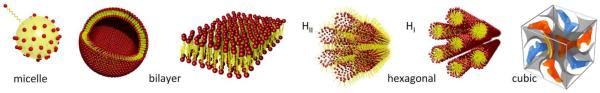
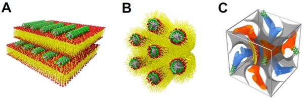
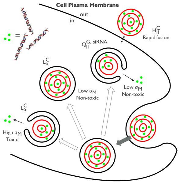
Lipids and nucleic acids (NAs) can hierarchically self-organize into a variety of nanostructures of increasingly complex geometries such as the 1D lamellar, 2D hexagonal, and 3D bicontinuous cubic phases. The diversity and complexity of those lipid-NA assemblies are interesting from a fundamental perspective as well as being relevant to the performance in gene delivery and gene silencing applications. The finding that not only the chemical make of the lipid-NA constructs, but their actual supramolecular organization, affects their gene transfection and silencing efficiencies has inspired physicists, chemists, and engineers to this field of research. At the moment it remains an open question how exactly the different lipid-NA structures interact with cells and organelles in order to output an optimal response. This article reviews our current understanding of the structures of different lipid-NA complexes and the corresponding cellular interaction mechanisms. The recent advances in designing optimal lipid-based NA carriers will be introduced with an emphasis on the structure-function relations.

Keywords: gene delivery; lipid-DNA; lipid-siRNA; self-assembly.


Figures

Figure 1
Figure 1
Schematic representation of various lipid self-assembled structures. Lipids in the presence of water can form micelle, liposome, inverse and normal hexagonal phases, bicontinuous cubic phase, respectively from left to right.
Figure 2
Figure 2
(A) The lamellar phase of CL-DNA complexes (LαC). DNA monolayers (green) are intercalated between lipid bilayers. (B) The inverted hexagonal phase of CL-DNA complexes (HIIC). DNA molecules (green) are inserted within water channels of the inverse lipid tubules forming a hexagonal array. (C) The unit cell of the double gyroid lipid cubic phase of CL-siRNA complexes (QIIG,siRNA). siRNAs (green) are incorporated within two intertwined but independent water channels (blue and orange). The lipid bilayer is represented as a gray surface.
Figure 3
Figure 3
Schematic illustration of (A) lipid-only and (B) lipid-siRNA Im3m cubic phase nanoparticles showing the internal bicontinuous cubic structure. siRNA (green) is incorporated in the water channels. PEG chains are located towards the outside of the particles as represented with gray tails. Cryo-Em images of (C) lipid-only and (D) lipid-siRNA Im3m cubic phase particulate systems obtained from GMO/DOTAP/GMO-PEG (95/4/1)-siRNA mixtures. Before and after siRNA incorporation, the internal cubic symmetry is preserved while maintaining the spherical shape of the particles. Note that water channels in (D) appear darker than that in (C) implying the presence of encapsulated siRNA molecules within the channels.
Figure 4
Figure 4
A schematic representation of the cellular pathway and transfection mechanisms of different CL-NA complexes. The lamellar phases of CL-DNA complexes (LαC) enter the cells through endocytosis while the inverted hexagonal phases of CL-DNA complexes (HIIC) rather directly interacts with the plasma membrane through facilitated fusion process. The fate of entered LαC complexes (i.e. trapped in endosomes) turned out to be dependent on the membrane charge density (σM). High σM facilitates the fusion with the endosomal membrane effectively releasing DNA inside the cell, but increases the cytotoxicity at the same time. Low σM involves the low cytotoxicity but this state lacks the capability to escape out of the endosomes. On the other hand, the cubic phase of CL-siRNA complexes (QIIG,siRNA) show the high efficiency of escaping endosomes even at low σM, which arises from the negative Gaussian curvatures intrinsic to bicontinuous cubic structures.

References

    1. Bruinsma R. Electrostatics of DNA-cationic lipid complexes: isoelectric instability. Eur. Phys. J. B. 1998;4:75–88.
    1. Golubović L, Golubović M. Fluctuations of Quasi-Two-Dimensional Smectics Intercalated between Membranes in Multilamellar Phases of DNA-Cationic Lipid Complexes. Phys. Rev. Lett. 1998;80:4341–4344.
    1. Harries D, May S, Gelbart WM, Ben-Shaul A. Structure, stability, and thermodynamics of lamellar DNA-lipid complexes. Biophys. J. 1998;75:159–173. - PMC - PubMed
    1. Gelbart WM, Bruinsma RF, Pincus PA, Parsegian VA. DNA-Inspired Electrostatics. Phys. Today. 2000;53:38.
    1. Evans DF, Wennerström H. The colloidal domain: where physics, chemistry, biology, and technology meet. Wiley-VCH; 1999.

LinkOut - more resources