. 2017 Apr 27:8:504.
doi: 10.3389/fimmu.2017.00504. eCollection 2017.

Inosine Released from Dying or Dead Cells Stimulates Cell Proliferation via Adenosine Receptors

Affiliations

Inosine Released from Dying or Dead Cells Stimulates Cell Proliferation via Adenosine Receptors

Jin Chen et al. Front Immunol. .

Abstract

Introduction: Many antitumor therapies induce apoptotic cell death in order to cause tumor regression. Paradoxically, apoptotic cells are also known to promote wound healing, cell proliferation, and tumor cell repopulation in multicellular organisms. We aimed to characterize the nature of the regenerative signals concentrated in the micromilieu of dead and dying cells.

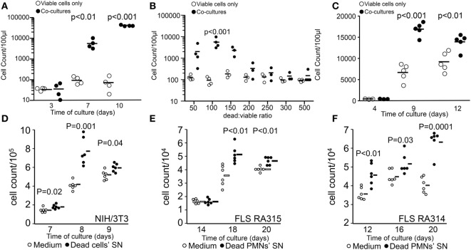
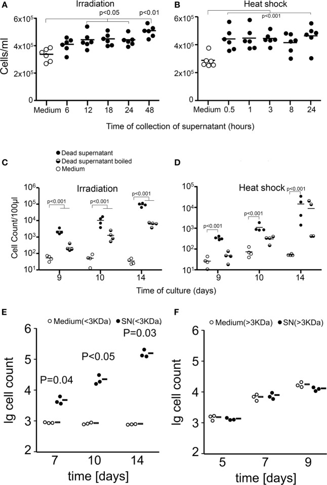
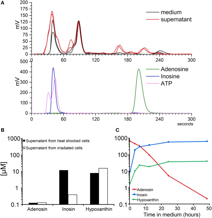
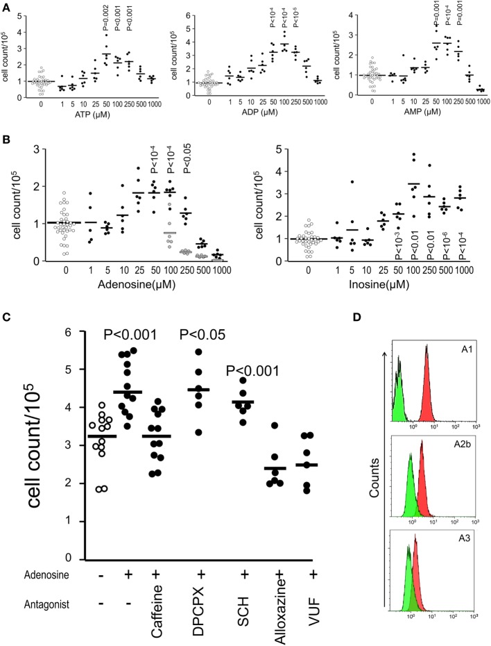
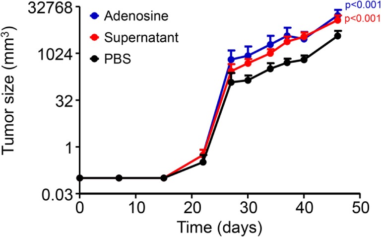
Methods: Cultures of viable melanoma B16F10 cells, mouse fibroblasts, and primary human fibroblast-like synoviocytes (FLS) in the presence of dead and dying cells, their supernatants (SNs), or purified agonists and antagonists were used to evaluate the stimulation of proliferation. Viable cell quantification was performed by either flow cytometry of harvested cells or by crystal violet staining of adherent cells. High-performance liquid chromatography and liquid chromatography coupled with mass spectrometry of cell SNs were deployed to identify the nature of growth-promoting factors. Coimplantation of living cells in the presence of SNs collected from dead and dying cells and specific agonists was used to evaluate tumor growth in vivo.

Results: The stimulation of proliferation of few surviving cells by bystander dead cells was confirmed for melanoma cells, mouse fibroblasts, and primary FLS. We found that small soluble molecules present in the protein-free fraction of SNs of dead and dying cells were responsible for the promotion of proliferation. The nucleoside inosine released by dead and dying cells acting via adenosine receptors was identified as putative inducer of proliferation of surviving tumor cells after irradiation and heat treatment.

Conclusion: Inosine released by dead and dying cells mediates tumor cell proliferation via purinergic receptors. Therapeutic strategies surmounting this pathway may help to reduce the rate of recurrence after radio- and chemotherapy.

Keywords: adenosine receptor; apoptosis; inosine; melanoma; necrosis; proliferation; repopulation.


Figures

Figure 1
Figure 1
Stimulation of tumor cell proliferation by bystander dying cells, the “feeder cells” effect. The upper panel shows viable cell counts of cocultures [closed circles in panels (A–C)] and viable cells alone in D10 medium [open circles in panels (A–C)]. Cocultures were composed of viable (200 cells/well) and lethally ultraviolet light type B-irradiated (10,000 cells/well) B16F10 melanoma cells (A). Dead/viable ratio titration of cocultures (B). Transwell cocultures with 10,000 apoptotic cells (C). The lower panel shows cell counts of independent wells containing fibroblasts in R10 medium [open circles in panels (D–F)] or in supernatants (SN) from apoptotic cells [closed circles in panels (D–F)]. Mouse NIH/3T3 fibroblast (1,000 cells/well) cultures with SNs of 0.2 million cells/ml apoptotic homologous cells (D). Human fibroblast-like synoviocytes (FLS, 1,000 cells/well) from two rheumatoid arthritis patients RA315 (E) and RA314 (F) cocultured with SNs from apoptotic neutrophils (0.2 million cells/ml).
Figure 2
Figure 2
Apoptotic and necrotic cells release proliferation-stimulating factors. Shown are cell counts of cultures of viable cells in medium alone (open circles) or in supernatants (SNs) from dead and dying cells (closed circles). Factors stimulating proliferation are released by dying and dead cells after 12 h of irradiation [apoptosis (A)] and immediately after heat shock [necrosis (B)]. Proliferation was measured by the crystal violet assay for adherent cells. Boiling of SNs (half-full circles) partially reduces but not abolishes the stimulation of proliferation of tumor cells [24 h after irradiation (C) and 1 h after heat shock (D)]. Factors stimulating the proliferation of tumor cells are present in the protein-free fraction (E) and not in the protein-rich fraction (F) of dead cell SNs.
Figure 3
Figure 3
Molecular analysis of the protein-free fraction of supernatants (SNs) from dying cells. Electrograms of SNs of B16F10 cells after 24 h of irradiation and metabolite standards by high-performance liquid chromatography (A). Concentrations of nucleosides measured by mass spectrometry of SNs of B16F10 cells after 24 h of irradiation and 1 h after heat shock (B). Spontaneous adenosine degradation into its metabolites inosine and hypoxanthine in SN spiked with 500 µM of adenosine and measured by mass spectrometry (C).
Figure 4
Figure 4
Adenosine receptors mediate the stimulation of the proliferation induced by supernatants of dead and dying cells. Shown are cell counts of cultures measured by the crystal violet method for adherent cells at day 9 showing the relative potency of nucleotides (A) and nucleosides (B) on the stimulation of proliferation of B16F10 melanoma cells (D10 medium wells are open circles, purinergic metabolites are closed circles, and vehicles for adenosine are gray circles). The pan-adenosine receptor antagonist caffeine and the specific adenosine receptor antagonists alloxazine (A2bR) and VUF (A3R) blocked the stimulation of proliferation induced by inosine (C). Adenosine receptor expression on B16F10 cells (D). Green represent the isotype antibody binding and red the receptor antibody.
Figure 5
Figure 5
Stimulation of proliferation in vivo. The presence of adenosine or supernatant of irradiated cells at the site of implantation of 50,000 B16F10 cells caused development of larger subcutaneous tumors in C57Bl/6 mice (n = 6 per group).

References

    1. Thompson CB. Apoptosis in the pathogenesis and treatment of disease. Science (1995) 267(5203):1456–62.10.1126/science.7878464 - DOI - PubMed
    1. Janko C, Munoz L, Chaurio R, Maueroder C, Berens C, Lauber K, et al. Navigation to the graveyard-induction of various pathways of necrosis and their classification by flow cytometry. Methods Mol Biol (2013) 1004:3–15.10.1007/978-1-62703-383-1_1 - DOI - PubMed
    1. Golstein P, Kroemer G. Cell death by necrosis: towards a molecular definition. Trends Biochem Sci (2007) 32(1):37–43.10.1016/j.tibs.2006.11.001 - DOI - PubMed
    1. Gregory CD, Pound JD. Cell death in the neighbourhood: direct microenvironmental effects of apoptosis in normal and neoplastic tissues. J Pathol (2011) 223(2):177–94.10.1002/path.2792 - DOI - PubMed
    1. Hochreiter-Hufford A, Ravichandran KS. Clearing the dead: apoptotic cell sensing, recognition, engulfment, and digestion. Cold Spring Harb Perspect Biol (2013) 5(1):a008748.10.1101/cshperspect.a008748 - DOI - PMC - PubMed