

Electrical Stimulation for Wound-Healing: Simulation on the Effect of Electrode Configurations
- PMID: 28497054
- PMCID: PMC5401728
- DOI: 10.1155/2017/5289041
Electrical Stimulation for Wound-Healing: Simulation on the Effect of Electrode Configurations
Abstract
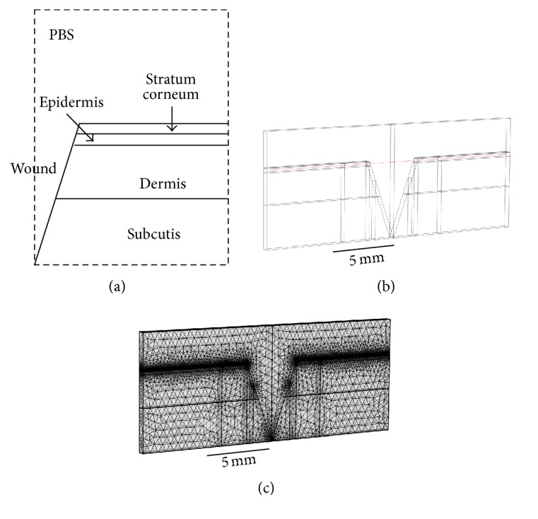
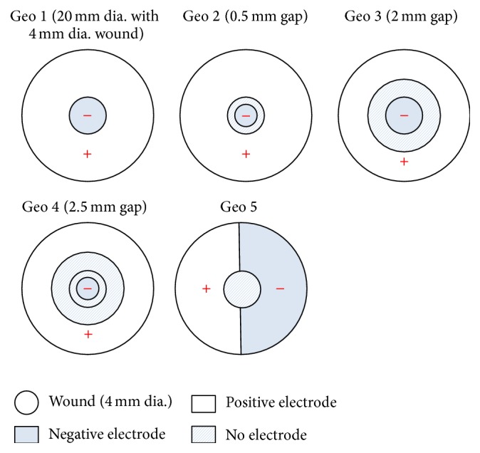
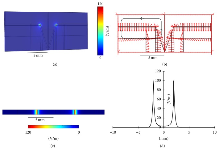
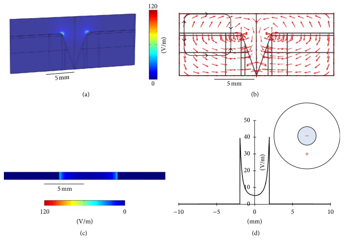
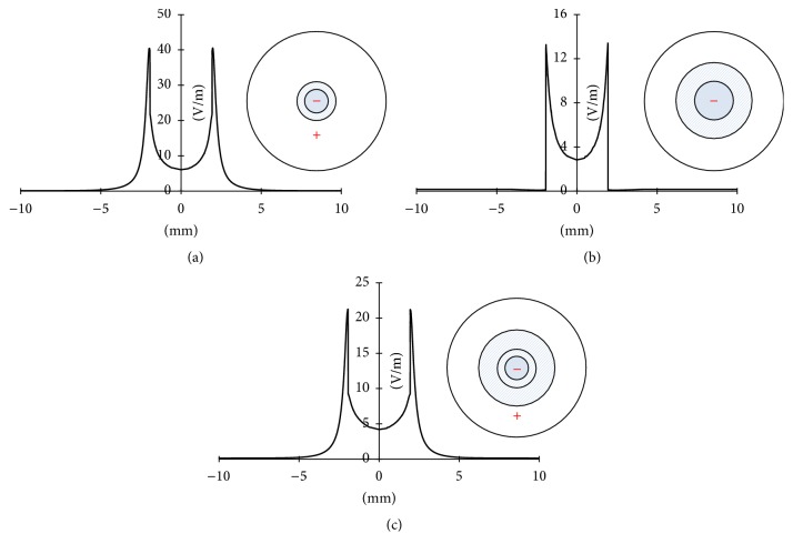
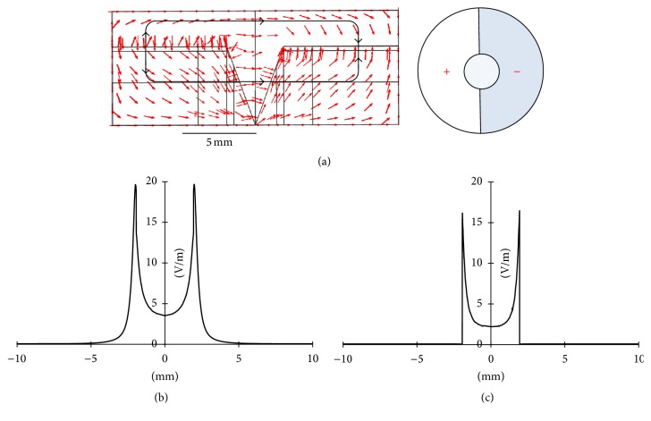
Endogenous electric field is known to play important roles in the wound-healing process, mainly through its effects on protein synthesis and cell migration. Many clinical studies have demonstrated that electrical stimulation (ES) with steady direct currents is beneficial to accelerating wound-healing, even though the underlying mechanisms remain unclear. In the present study, a three-dimensional finite element wound model was built to optimize the electrode configuration in ES. Four layers of the skin, stratum corneum, epidermis, dermis, and subcutis, with defined thickness and electrical properties were modeled. The main goal was to evaluate the distributions of exogenous electric fields delivered with direct current (DC) stimulation using different electrode configurations such as sizes and positions. Based on the results, some guidelines were obtained in designing the electrode configuration for applications of clinical ES.
Figures
Similar articles
-
A new electrode design to improve outcomes in the treatment of chronic non-healing wounds in diabetes.Diabetes Technol Ther. 2009 May;11(5):315-22. doi: 10.1089/dia.2008.0092. Diabetes Technol Ther. 2009. PMID: 19425879
-
The combined effect of a three-channel electrode delivery system with local heat on the healing of chronic wounds.Diabetes Technol Ther. 2009 Oct;11(10):681-8. doi: 10.1089/dia.2009.0024. Diabetes Technol Ther. 2009. PMID: 19821762
-
In vivo 3-D distributions of electric fields in pig skin with rectangular pulse electrical current stimulation (RPECS).Bioelectromagnetics. 1996;17(4):253-62. doi: 10.1002/(SICI)1521-186X(1996)17:4<253::AID-BEM1>3.0.CO;2-2. Bioelectromagnetics. 1996. PMID: 8891184
-
Electrical stimulation for pressure sore prevention and wound healing.Assist Technol. 2000;12(1):50-66. doi: 10.1080/10400435.2000.10132009. Assist Technol. 2000. PMID: 11067577 Review.
-
Accelerated Skin Wound Healing by Electrical Stimulation.Adv Healthc Mater. 2021 Aug;10(16):e2100557. doi: 10.1002/adhm.202100557. Epub 2021 May 4. Adv Healthc Mater. 2021. PMID: 33945225 Review.
Cited by
-
Microcurrent Stimulation Triggers MAPK Signaling and TGF-β1 Release in Fibroblast and Osteoblast-Like Cell Lines.Cells. 2020 Aug 19;9(9):1924. doi: 10.3390/cells9091924. Cells. 2020. PMID: 32825091 Free PMC article.
-
Propagating acoustic waves on a culture substrate regulate the directional collective cell migration.Microsyst Nanoeng. 2021 Nov 11;7:90. doi: 10.1038/s41378-021-00304-8. eCollection 2021. Microsyst Nanoeng. 2021. PMID: 34786204 Free PMC article.
-
Wearable electronics for skin wound monitoring and healing.Soft Sci. 2022;2:9. doi: 10.20517/ss.2022.13. Epub 2022 Jun 30. Soft Sci. 2022. PMID: 37056725 Free PMC article.
-
CD9 negatively regulates collective electrotaxis of the epidermal monolayer by controlling and coordinating the polarization of leader cells.Burns Trauma. 2023 Jul 24;11:tkad012. doi: 10.1093/burnst/tkad012. eCollection 2023. Burns Trauma. 2023. PMID: 37492637 Free PMC article.
-
Changes in the extracellular microenvironment and osteogenic responses of mesenchymal stem/stromal cells induced by in vitro direct electrical stimulation.J Tissue Eng. 2021 Feb 16;12:2041731420974147. doi: 10.1177/2041731420974147. eCollection 2021 Jan-Dec. J Tissue Eng. 2021. PMID: 33643602 Free PMC article.
References
MeSH terms
LinkOut - more resources
Full Text Sources
Other Literature Sources
