. 2017 Apr 19:5:165-179.
doi: 10.1016/j.omtm.2017.04.005. eCollection 2017 Jun 16.

Efficient Production of Papillomavirus Gene Delivery Vectors in Defined In Vitro Reactions

Affiliations

Efficient Production of Papillomavirus Gene Delivery Vectors in Defined In Vitro Reactions

Carla Cerqueira et al. Mol Ther Methods Clin Dev. .

Abstract

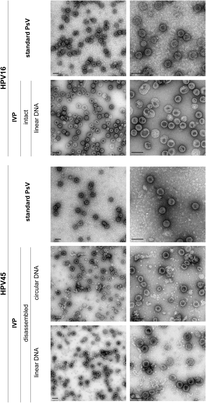
Papillomavirus capsids can package a wide variety of nonviral DNA plasmids and deliver the packaged genetic material to cells, making them attractive candidates for targeted gene delivery vehicles. However, the papillomavirus vectors generated by current methods are unlikely to be suitable for clinical applications. We have developed a chemically defined, cell-free, papillomavirus-based vector production system that allows the incorporation of purified plasmid DNA (pseudogenome) into high-titer papillomavirus L1/L2 capsids. We investigated the incorporation of several DNA forms into a variety of different papillomavirus types, including human and animal types. Our results show that papillomavirus capsids can package and transduce linear or circular DNA under defined conditions. Packaging and transduction efficiencies were surprisingly variable across capsid types, DNA forms, and assembly reaction conditions. The pseudoviruses produced by these methods are sensitive to the same entry inhibitors as cell-derived pseudovirions, including neutralizing antibodies and heparin. The papillomavirus vector production systems developed in this study generated as high as 1011 infectious units/mg of L1. The pseudoviruses were infectious both in vitro and in vivo and should be compatible with good manufacturing practice (GMP) requirements.

Keywords: gene delivery; gene packaging; papillomavirus.


Figures

Figure 1
Figure 1
HPV16 Can Package Linear DNA Intact or disassembled HPV16 VLPs were reassembled with a GFP reporter plasmid with (+) or without (−) nuclear extract as indicated. Three types of GFP reporter plasmids were used: circular supercoiled, linearized, or blunt (lacking the potential to be re-circularized). After reassembly and nuclease treatment, the resultant PsVs were used to infect HeLa cells. The number of infected cells (GFP-positive) was determined 72 hr p.i. by flow cytometric analysis. Shown is a representative experiment. The error bars represent the deviation between duplicates.
Figure 2
Figure 2
Disassembly of Different PV VLPs HPV16, HPV45, or HPV2 particles were disassembled in 100 mM NaCl, 20 mM Tris (pH 8.2), 2 mM DTT, and 0.01% Tween 80 for 3 hr at 37°C. BPV1 particles were disassembled in 50 mM NaCl, 20 mM Tris (pH 8.2), 2 mM DTT, and 0.01% Tween 80 for 3 hr at 37°C. Samples were analyzed by transmission electron microscopy. Scale bars represent 100 nm.
Figure 3
Figure 3
Optimization of In Vitro Reassembly (A) Reassembly of intact HPV16 or disassembled HPV45 was assessed. Reassembly reactions were performed at the indicated pH and NaCl concentrations for 20 hr at 37°C with 150 ng of GFP plasmid (either circular or linearized plasmid as indicated). All samples were nuclease-treated prior to infection of HeLa cells. Infection was scored 72 hr p.i. Shown is a representative experiment. The error bars show the deviation between duplicates. (B) Intact HPV16 was incubated at pH 5.2 with the indicated amounts of linearized GFP plasmid (linear DNA). Disassembled HPV45 was incubated at pH 7.2 and 150 mM NaCl with the indicated amounts of circular or linearized GFP plasmid. Reactions were incubated 20 hr at 37°C and then nuclease-treated prior to infection. Shown are representative experiments. The error bars show the deviation between duplicates.
Figure 4
Figure 4
Electron Microscopy of HPV16 and HPV45 IVP Vectors Intact HPV16 particles were assembled at pH 5.2 with a linearized Luc/GFP plasmid for 30 hr at 37°C. HPV45 was disassembled and then reassembled at pH 7.2 with 150 mM NaCl, 10 mM CaCl2, and 0.02% Tween 80 with linearized or circular Luc/GFP for 30 hr at 37°C. Both HPV16 and HPV45 were then incubated for a further 15 hr with 5 mM GSSG. After nuclease treatment, samples were centrifuged over an Optiprep cushion, and the virus-containing fraction was analyzed by electron microscopy. HPV16 or HPV45 PsVs produced by the standard cell-based protocol were included for comparison. Scale bars represent 100 nm.
Figure 5
Figure 5
PsVs Prepared by the IVP Method Have Similar Neutralization and Biochemical Inhibition Profiles as Standard PsVs (A and B) Standard or HPV16 IVP PsVs containing a packaged Luc/GFP plasmid were pre-incubated with dilutions of heparin (A) or H16.V5 antibody (B) for 1 hr on ice prior to infection of 293TT cells. The percentage of infected cells (GFP-positive) was determined 72 hr p.i. by flow cytometry. Shown are the mean values for at least three independent experiments ± SD normalized to untreated virus. (C and D) Standard or IVP HPV45 PsVs were pre-incubated with dilutions of heparin (C) or polyclonal HPV45 serum (D) for 1 hr on ice. Infection and analysis were performed as for (A) and (B). (E and F) 293TT cells were infected with standard or IVP PsVs in the presence of 10 μM furin inhibitor (dec-RVKR-cmk), 20 mM NH4Cl, 300 nM γ-secretase inhibitor (compound XXI), or 10 μM CsA, or left untreated. The percentage of infected cells (GFP-positive) was determined 72 hr p.i. by flow cytometry. (E) Infection with HPV16. (F) Infection with HPV45. Shown are the mean values for at least three independent experiments ± SD normalized for untreated cells. The physical state of the packaged plasmid is indicated as either circular or linear.
Figure 6
Figure 6
Kinetics of In Vivo Intravaginal Infection (A–H) Depo-Provera-treated BALB/c mice were nonoxynol-9-treated prior to infection. 1 × 107 infectious units HPV16 (A–C), HPV45 (D–F), and HPV58 (G) or 3 × 106 infectious units HPV26 (H) containing a packaged Luc/GFP plasmid were inoculated intravaginally (5 mice/group). The plasmid was either circular, linear, or linear split as indicated. Luciferase expression was monitored daily following delivery. The mean value of the average radiance ± SEM measurement per group is shown. Three different virus preparations for each condition, as indicated, were compared with infection by standard PsV preparations. Note the different scale for the HPV26 experiment.

References

    1. Buck C.B., Cheng N., Thompson C.D., Lowy D.R., Steven A.C., Schiller J.T., Trus B.L. Arrangement of L2 within the papillomavirus capsid. J. Virol. 2008;82:5190–5197. - PMC - PubMed
    1. Guan J., Bywaters S.M., Brendle S.A., Ashley R.E., Makhov A.M., Conway J.F., Christensen N.D., Hafenstein S. Cryoelectron microscopy maps of human papillomavirus 16 reveal L2 densities and heparin binding site. Structure. 2016;25:253–263. - PubMed
    1. Chen H.S., Conway M.J., Christensen N.D., Alam S., Meyers C. Papillomavirus capsid proteins mutually impact structure. Virology. 2011;412:378–383. - PMC - PubMed
    1. Ishii Y., Ozaki S., Tanaka K., Kanda T. Human papillomavirus 16 minor capsid protein L2 helps capsomeres assemble independently of intercapsomeric disulfide bonding. Virus Genes. 2005;31:321–328. - PubMed
    1. Day P.M., Roden R.B., Lowy D.R., Schiller J.T. The papillomavirus minor capsid protein, L2, induces localization of the major capsid protein, L1, and the viral transcription/replication protein, E2, to PML oncogenic domains. J. Virol. 1998;72:142–150. - PMC - PubMed