

CHARMM-GUI ligand reader and modeler for CHARMM force field generation of small molecules
- PMID: 28497616
- PMCID: PMC5488718
- DOI: 10.1002/jcc.24829
CHARMM-GUI ligand reader and modeler for CHARMM force field generation of small molecules
Abstract
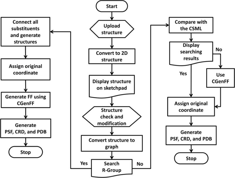
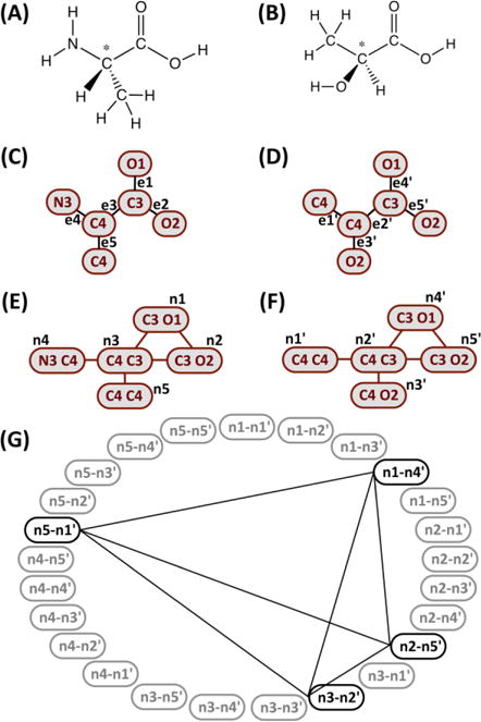
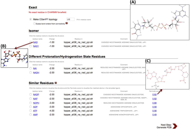
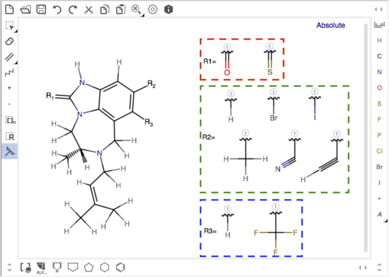
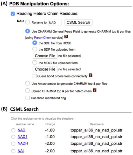
Reading ligand structures into any simulation program is often nontrivial and time consuming, especially when the force field parameters and/or structure files of the corresponding molecules are not available. To address this problem, we have developed Ligand Reader & Modeler in CHARMM-GUI. Users can upload ligand structure information in various forms (using PDB ID, ligand ID, SMILES, MOL/MOL2/SDF file, or PDB/mmCIF file), and the uploaded structure is displayed on a sketchpad for verification and further modification. Based on the displayed structure, Ligand Reader & Modeler generates the ligand force field parameters and necessary structure files by searching for the ligand in the CHARMM force field library or using the CHARMM general force field (CGenFF). In addition, users can define chemical substitution sites and draw substituents in each site on the sketchpad to generate a set of combinatorial structure files and corresponding force field parameters for throughput or alchemical free energy simulations. Finally, the output from Ligand Reader & Modeler can be used in other CHARMM-GUI modules to build a protein-ligand simulation system for all supported simulation programs, such as CHARMM, NAMD, GROMACS, AMBER, GENESIS, LAMMPS, Desmond, OpenMM, and CHARMM/OpenMM. Ligand Reader & Modeler is available as a functional module of CHARMM-GUI at http://www.charmm-gui.org/input/ligandrm. © 2017 Wiley Periodicals, Inc.
Keywords: CGenFF; drug design; molecular dynamics simulation; molecular modeling; protein-ligand interactions.
© 2017 Wiley Periodicals, Inc.
Figures

Similar articles
-
CHARMM-GUI 10 years for biomolecular modeling and simulation.J Comput Chem. 2017 Jun 5;38(15):1114-1124. doi: 10.1002/jcc.24660. Epub 2016 Nov 14. J Comput Chem. 2017. PMID: 27862047 Free PMC article. Review.
-
CHARMM-GUI Glycan Modeler for modeling and simulation of carbohydrates and glycoconjugates.Glycobiology. 2019 Apr 1;29(4):320-331. doi: 10.1093/glycob/cwz003. Glycobiology. 2019. PMID: 30689864 Free PMC article.
-
Glycan Reader is improved to recognize most sugar types and chemical modifications in the Protein Data Bank.Bioinformatics. 2017 Oct 1;33(19):3051-3057. doi: 10.1093/bioinformatics/btx358. Bioinformatics. 2017. PMID: 28582506 Free PMC article.
-
CHARMM-GUI Input Generator for NAMD, GROMACS, AMBER, OpenMM, and CHARMM/OpenMM Simulations Using the CHARMM36 Additive Force Field.J Chem Theory Comput. 2016 Jan 12;12(1):405-13. doi: 10.1021/acs.jctc.5b00935. Epub 2015 Dec 3. J Chem Theory Comput. 2016. PMID: 26631602 Free PMC article.
-
CHARMM-GUI PDB manipulator for advanced modeling and simulations of proteins containing nonstandard residues.Adv Protein Chem Struct Biol. 2014;96:235-65. doi: 10.1016/bs.apcsb.2014.06.002. Epub 2014 Aug 24. Adv Protein Chem Struct Biol. 2014. PMID: 25443960 Free PMC article. Review.
Cited by
-
Inhibition of SARS-CoV-2 main protease: a repurposing study that targets the dimer interface of the protein.J Biomol Struct Dyn. 2022 Sep;40(15):7167-7182. doi: 10.1080/07391102.2021.1910571. Epub 2021 Apr 13. J Biomol Struct Dyn. 2022. PMID: 33847241 Free PMC article.
-
Ionizable Cationic Lipids and Helper Lipids Synergistically Contribute to RNA Packing and Protection in Lipid-Based Nanomaterials.J Phys Chem B. 2024 Oct 17;128(41):10165-10177. doi: 10.1021/acs.jpcb.4c05057. Epub 2024 Oct 4. J Phys Chem B. 2024. PMID: 39366669
-
CHARMM-GUI Multicomponent Assembler for modeling and simulation of complex multicomponent systems.Nat Commun. 2024 Jun 27;15(1):5459. doi: 10.1038/s41467-024-49700-4. Nat Commun. 2024. PMID: 38937468 Free PMC article.
-
In silico identification of widely used and well-tolerated drugs as potential SARS-CoV-2 3C-like protease and viral RNA-dependent RNA polymerase inhibitors for direct use in clinical trials.J Biomol Struct Dyn. 2021 Oct;39(17):6772-6791. doi: 10.1080/07391102.2020.1802346. Epub 2020 Aug 5. J Biomol Struct Dyn. 2021. PMID: 32752938 Free PMC article.
-
Supramolecular organization and dynamics of mannosylated phosphatidylinositol lipids in the mycobacterial plasma membrane.Proc Natl Acad Sci U S A. 2023 Jan 31;120(5):e2212755120. doi: 10.1073/pnas.2212755120. Epub 2023 Jan 24. Proc Natl Acad Sci U S A. 2023. PMID: 36693100 Free PMC article.
References
-
- MacKerell AD, Bashford D, Bellott M, Dunbrack RL, Evanseck JD, Field MJ, Fischer S, Gao J, Guo H, Ha S, Joseph-McCarthy D, Kuchnir L, Kuczera K, Lau FT, Mattos C, Michnick S, Ngo T, Nguyen DT, Prodhom B, Reiher WE, Roux B, Schlenkrich M, Smith JC, Stote R, Straub J, Watanabe M, Wiorkiewicz-Kuczera J, Yin D, Karplus M. J Phys Chem B. 1998;102(18):3586–3616. - PubMed
-
- Oostenbrink C, Villa A, Mark AE, van Gunsteren WF. J Comput Chem. 2004;25(13):1656–1676. - PubMed
-
- Cornell WD, Cieplak P, Bayly CI, Gould IR, Merz KM, Ferguson DM, Spellmeyer DC, Fox T, Caldwell JW, Kollman PA. J Am Chem Soc. 1995;117(19):5179–5197.
-
- Jorgensen WL, Maxwell DS, TiradoRives J. J Am Chem Soc. 1996;118(45):11225–11236.
-
- Rappe AK, Casewit CJ, Colwell KS, Goddard WA, Skiff WM. J Am Chem Soc. 1992;114(25):10024–10035.
Publication types
Grants and funding
LinkOut - more resources
Full Text Sources
Other Literature Sources
