. 2017 May 12:8:15231.
doi: 10.1038/ncomms15231.

Structural heterogeneity in the intrinsically disordered RNA polymerase II C-terminal domain

Affiliations

Structural heterogeneity in the intrinsically disordered RNA polymerase II C-terminal domain

Bede Portz et al. Nat Commun. .

Abstract

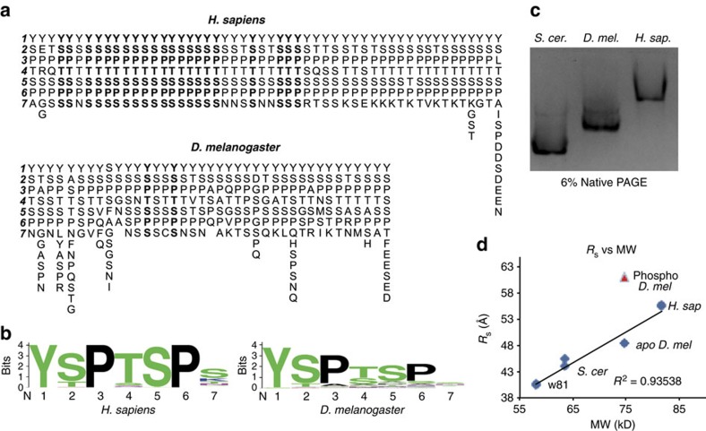
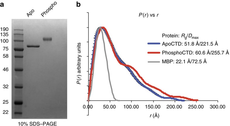
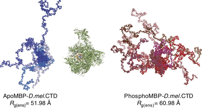
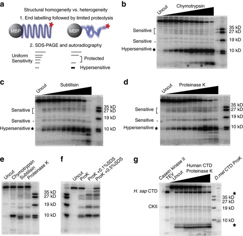
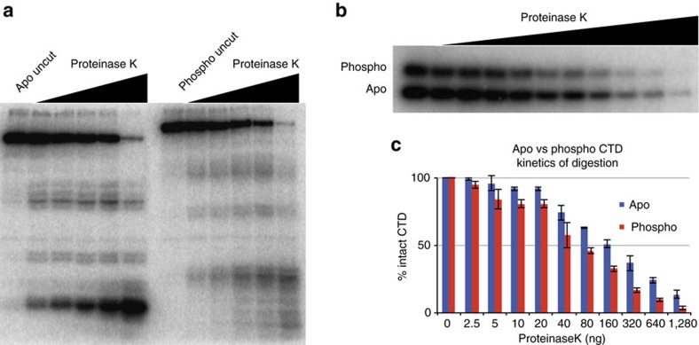
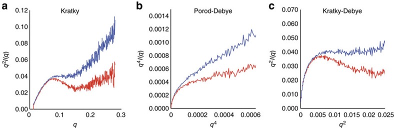
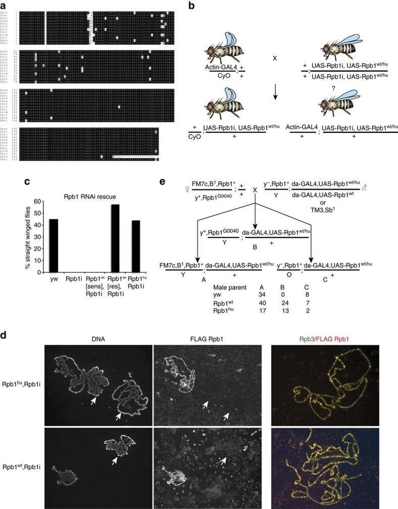
RNA polymerase II contains a repetitive, intrinsically disordered, C-terminal domain (CTD) composed of heptads of the consensus sequence YSPTSPS. The CTD is heavily phosphorylated and serves as a scaffold, interacting with factors involved in transcription initiation, elongation and termination, RNA processing and chromatin modification. Despite being a nexus of eukaryotic gene regulation, the structure of the CTD and the structural implications of phosphorylation are poorly understood. Here we present a biophysical and biochemical interrogation of the structure of the full length CTD of Drosophila melanogaster, which we conclude is a compact random coil. Surprisingly, we find that the repetitive CTD is structurally heterogeneous. Phosphorylation causes increases in radius, protein accessibility and stiffness, without disrupting local structural heterogeneity. Additionally, we show the human CTD is also structurally heterogeneous and able to substitute for the D. melanogaster CTD in supporting fly development to adulthood. This finding implicates conserved structural organization, not a precise array of heptad motifs, as important to CTD function.


Conflict of interest statement

The authors declare no competing financial interests.

Figures

Figure 1
Figure 1. A similar global CTD structure exists despite sequence differences.
(a) The sequences of the CTDs of D. melanogaster and H. sapiens with each repeat beginning with YSP arranged vertically and the CTD oriented from left to right. Consensus heptads are in boldface. The Drosophila CTD has only two consensus heptads. (b) Motif conservation of fly and human CTDs. (c) Native gel electrophoresis of the S. cerevisae, D. melanogaster and H. sapiens MBP-CTD fusion proteins, with pIs of 5.57, 5.83, 5.93, respectively. Electrophoretic mobility scales with CTD length, suggesting structural homology. (d) RS versus molecular weight (MW) derived from size exclusion chromatography analysis of MBP-CTD fusions. Two replicates of each protein are plotted (several points appear as one because of overlapping values). RS is linearly related to MW, suggesting gross structural homology. Phosphorylation of the D. melanogaster CTD by P-TEFb increases RS, causing the CTD to deviate from the line (red triangles).
Figure 2
Figure 2. The CTD extends as a function of phosphorylation.
(a) Coomassie blue stained 12% SDS–PAGE gel of 600 ng of apo or phospho MBP-D.melCTD used for SAXS experiments. (b) Pairwise distance distribution (P(r) versus r) plots of the apo (blue line) and phospho (red line) MBP-D.melCTD, and the CRYSOL generated scattering curve of MBP (grey line). The phospho CTD adopts a more extended conformation compared to the apo CTD, with increases in Rg and Dmax.
Figure 3
Figure 3. Ensemble optimization models depicting CTD extension.
Averaged apo and phospho MBP-D.melCTD scattering data were modelled using EOM 2.1 from the ATSAS suite. Individual models in the apo and phospho ensembles are shown in shades of blue and red, respectively, oriented by MBP and positioned adjacent to the 12 subunit RNA Pol II elongation complex (1Y1W.pdb) model (green) for scale.
Figure 4
Figure 4. The CTD is structurally heterogeneous across its length.
(a) Experimental design: end-labelled MBP-D.melCTD is subject to limited proteolysis, SDS-PAGE, and autoradiography. A completely unstructured, or alternatively, a structurally repetitive CTD is predicted to generate a uniform pattern of proteolytic fragments. A structurally heterogeneous CTD is predicted to give rise to a non-uniform pattern. (b) Limited proteolysis with chymotrypsin generates a non-uniform pattern of CTD fragments, with a hypersensitive site in the distal CTD, a sensitive region in the proximal CTD, and a largely protease insensitive region in the central CTD that is cleaved at only one site. The right most lane contains radiolabelled CTD fusion proteins with the molecular weights designated on the right of the panel. (c,d) Limited proteolysis with subtilisin or proteinase K reveal similar sites of sensitivity and protection. (e) A direct comparison of proteolytic fragments generated by three proteases shows a similar by not identical pattern of proteolysis. (f) Sodium dodecyl sulfate (SDS) alters the relative proteolytic sensitivity of the CTD to proteinase K, enhancing the sensitivity of the CTD at sites that generate bands near 35 kD and below 19 kD relative to the CTD ladder (0.1% SDS lane). 0.5% SDS renders the globular MBP portion of the fusion protein more susceptible to proteolysis, evidenced by the proteolytic fragment above the 35 kD CTD ladder band but below the intact fusion. (g) The human CTD is radiolabelled on the final acidic repeat by casein kinase II (CK2) (uncut lane). TEV cleavage to separate the human CTD from the MBP fusion demarcates the point below which protease sensitivity occurs in the CTD portion of the protein (TEV lane). Limited proteolysis with proteinase K reveals a distal hypersensitive site reminiscent of that observed in the Drosophila CTD (compare to D.melCTD ProK lane), a protease hypersensitive proximal site near MBP and a central region that is largely protease insensitive. Both the human and fly CTDs share distal protease hypersensitivity, proximal sensitivity that is localized to a discrete region in the human CTD, and a more central region of the CTD that is less protease sensitive.
Figure 5
Figure 5. Local structural heterogeneity is maintained in the phospho CTD but the phospho CTD is more accessible to protein interaction.
(a) Limited proteolysis of the phospho CTD shows an altered pattern of proteolysis. The hypersensitivity of the distal site is reduced, and the pattern of proteolysis is more evenly distributed among the sensitive sites. The proximal sensitive region, and central protected region are preserved after phosphorylation, with proteolytic fragments shifted relative to the apo fragments due to phosphorylation across the CTD. (b) Representative limited proteolysis of a mixture of apo and phospho CTD to compare protease accessibility. (c) Average of three replicates of the apo (blue bars) and phospho (red bars) mixing experiments quantified as the percentage of intact CTD remaining at each protease concentration. 100–50% intact CTD is the single hit kinetics range of the experiment. Error bars depict s.e.m.
Figure 6
Figure 6. Phosphorylation stiffens the CTD.
(a) Kratky Plot (q2 × I(q) versus q) of averaged apo (blue) and phospho (red) MBP-CTD scattering curves. The more gradual rise in the high q region of the plot for the phospho-CTD than the apo-CTD indicates that the phospho-CTD is less flexible than the apo CTD. (b,c) Porod–Debye (q4 × I(q) versus q4) and Kratky Debye (q2 × I(q) versus q2) plots of the apo (blue) and phospho (red) scattering curves. The increased rise in the Porod-Debye plot for the apo-CTD compared to phospho-CTD indicates a phosphorylation dependent decrease in flexibility, as does the loss of the plateau in the Kratky–Debye plot for the phospho-CTD.
Figure 7
Figure 7. The human CTD can function in place of the Drosophila CTD in vivo.
(a) CTD amino acid sequence alignment from 12 species of Drosophila. (b) Experimental design of the human CTD rescue experiment. UAS-Rpb1i is a GAL4 inducible transgene that produces an RNAi against endogenous Rpb1. UAS-Rpb1 is a GAL4 inducible transgene that encodes a FLAG-tagged, RNAi-resistant derivative of Rpb1. Fly images were created with Genotype Builder, (c) Results of crosses to test if ectopically expressed derivatives of Rpb1 rescue the lethality caused by ubiquitously expressing Rpb1 RNAi. Each bar on the histogram indicates the percentage of progeny with straight wings. yw: ActGAL4/CyO × yw (n=137 progeny); Rpb1i: ActGAL4/CyO × UAS-Rpb1i (n=86 progeny); Rpb1wt[sens],Rpb1i: ActGAL4/CyO × UAS-Rpb1i,UAS-Rpb1sen (n=102 progeny); Rpb1wt[res],Rpb1i: ActGAL4/CyO × UAS-Rpb1i,UAS-Rpb1res (n=91 progeny); Rpb1hu,Rpb1i: ActGAL4/CyO × UAS-Rpb1i, UAS-Rpb1hu (n=78 progeny). (d) Immunofluorescence of polytene chromosomes from salivary glands of third instar larvae. Larvae were derived from mating ActGAL4/CyO and UAS-Rpb1i, UAS-Rpb1 parents, so half of the larvae ectopically expressed Drosophila or humanized Rpb1 (Left and Middle panels). FLAG Rpb1 detected on chromosomes co-localizes with the Rpb3 subunit of Pol II (Right panel). Arrows highlight chromosomes from individuals not expressing FLAG Rpb1 variants (CyO/+; UAS-Rpb1i,UAS-Rpb1/+). (e) Genetic complementation assay mating scheme and results. Complementation was scored in males, which have a single copy of the X chromosome on which the endogenous Rpb1 gene resides. Female flies carrying the early embryonic lethal Rpb1 mutant allele, G0040 are crossed to male flies expressing Rpb1wt or Rpb1hu under the control of da-GAL4, or to yw control flies. Crosses were carried out in triplicate with 8 males and 8 female parents per cross and flies were grown at 24C and 70% humidity. Shown in e are the total counts of male progeny; counts from individual vials are shown in Supplementary Table 1. ‘B' males indicate that the ectopically expressed Rpb1 complements of the lethal allele. We observe no ‘B' flies from the yw control crosses.

Similar articles

Cited by

References

    1. Chapman R. D., Heidemann M., Hintermair C. & Eick D. Molecular evolution of the RNA polymerase II CTD. Trends Genet. 24, 289–296 (2008). - PubMed
    1. Corden J. L. RNA polymerase II C-terminal domain: Tethering transcription to transcript and template. Chem. Rev. 113, 8423–8455 (2013). - PMC - PubMed
    1. Eick D. & Geyer M. The RNA polymerase II carboxy-terminal domain (CTD) code. Chem. Rev. 113, 8456–8490 (2013). - PubMed
    1. Meinhart A. & Cramer P. Recognition of RNA polymerase II carboxy-terminal domain by 3′-RNA-processing factors. Nature 430, 223–226 (2004). - PubMed
    1. Cagas P. M. & Corden J. L. Structural studies of a synthetic peptide derived from the carboxyl-terminal domain of RNA polymerase II. Proteins 21, 149–160 (1995). - PubMed

Publication types

MeSH terms

Substances

LinkOut - more resources