

Review
doi: 10.5152/dir.2017.16484.
Imaging for abdominal involvement in amyloidosis
Affiliations
- PMID: 28498108
- PMCID: PMC5508951
- DOI: 10.5152/dir.2017.16484
Item in Clipboard
Review
Imaging for abdominal involvement in amyloidosis
Diagn Interv Radiol.
2017 Jul-Aug.
Abstract
Involvement of the abdominal organs has variable presentations mostly without specific findings. The objective of this pictorial essay was to illustrate the computed tomography and magnetic resonance imaging (MRI) findings of abdominal involvement in systemic amyloidosis. Heterogeneous appearance of the liver, periportal involvement, diffuse low signal intensity of spleen on T2-weighted MRI, and thickened bowel wall may be helpful imaging findings when accompanied by presence or history of chronic inflammatory disease and clinical suspicion for amyloidosis.
Conflict of interest statement
The authors declared no conflicts of interest.
Figures

A 59-year-old woman with primary amyloidosis. Ultrasonography (a) shows diffuse heterogeneous liver parenchyma. Axial contrast-enhanced T1-weighted images (b, c) demonstrate diffuse heterogeneous liver parenchyma with periportal involvement (c,
black arrow). Note also diffuse irregular wall thickening of stomach (white arrow). Axial contrast-enhanced CT images (d, e) show periportal involvement (black arrows) and diffuse wall thickening of stomach (white arrow). In panel (f), microscopic finding (hematoxylin and eosin stain [H–E]; original magnification, × 400) reveals acute amorphous amyloid deposition in the portal area (arrows). Panel (g) shows immunohistochemical staining (× 400) of amorphous depositions in liver.

A 27-year-old man with familial Mediterranean fever. Axial T2-weighted image (a) demonstrates heterogeneous liver parenchyma. After one year, axial contrast-enhanced CT images (b, c) demonstrate infarct areas in the liver. Note also enlargement in the right adrenal gland (c,
arrow). In panel (d), microscopic finding (H–E stain; original magnification, × 400) reveals acute amorphous amyloid deposition around blood vessels in the portal area (arrows).

A 9-year-old girl with systemic amyloidosis. Axial T2-weighted images demonstrate (a) diffuse low signal intensity of liver and spleen, (b) enlargement of kidneys, (c) wall thickening of small intestine (arrows), (d) wall thickening of rectosigmoid colon (white arrow) and infiltration of perirectal fat plane (black arrow).

A 38-year-old man with primary amyloidosis. Axial contrast-enhanced CT images show (a) heterogeneous attenuation of spleen, (b) ascites (asterisk) and peritoneal involvement (arrow).
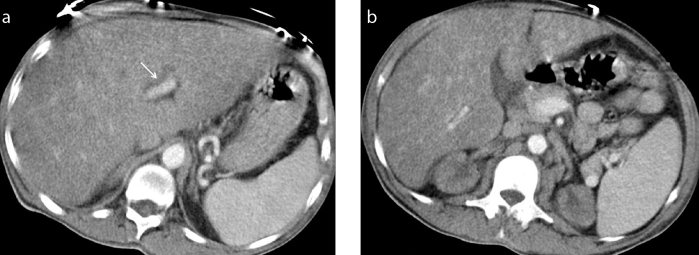
A 46-year-old man with familial Mediterranean fever. Axial contrast-enhanced CT images show (a) heterogeneous attenuation of liver and periportal involvement (arrow) and (b) bilateral small kidneys.

A 61-year-old man with familial Mediterranean fever. Axial contrast-enhanced CT images (a, b) show left renal pelvis involvement (a,
arrow) and diffuse bladder wall thickening (b,
arrow). Panel (c) shows immunohistochemical staining (× 400) of amorphous depositions in the colonic wall.
Comment in
-
T1 mapping and magnetic resonance elastography: potential new techniques for quantification of parenchymal changes in hepatic amyloidosis.Diagn Interv Radiol. 2017 Nov-Dec;23(6):478. doi: 10.5152/dir.2017.17240. Diagn Interv Radiol. 2017. PMID: 29097350 Free PMC article. No abstract available.
References
-
- Glenner GG. Amyloid deposits and amyloidosis. The beta-fibrilloses (first of two parts) N Engl J Med. 1980;302:1283–1292. - PubMed
-
- Monzawa S, Tsukamoto T, Omata K, Hosoda K, Araki T, Sugimura K. A case with primary amyloidosis of the liver and spleen: radiologic findings. Eur J Radiol. 2002;41:237–241. https://doi.org/10.1016/S0720-048X(01)00407-7. - DOI - PubMed
-
- Scott PP, Scott WW, Jr, Siegelman SS. Amyloidosis: an overview. Semin Roentgenol. 1986;21:103–112. https://doi.org/10.1016/0037-198X(86)90027-1. - DOI - PubMed
-
- Kyle RA, Bayrd ED. Amyloidosis: review of 236 cases. Medicine. 1975;54:271–299. https://doi.org/10.1097/00005792-197507000-00001. - DOI - PubMed
-
- Kim SH, Han JK, Lee KH, et al. Abdominal amyloidosis: spectrum of radiological findings. Clin Radiol. 2003;58:610–620. https://doi.org/10.1016/S0009-9260(03)00142-9. - DOI - PubMed
Publication types
MeSH terms
LinkOut - more resources
Full Text Sources
Other Literature Sources
Medical
