Review
. 2017 Jul;24(7):1184-1195.
doi: 10.1038/cdd.2017.65. Epub 2017 May 12.

Initiation and execution mechanisms of necroptosis: an overview

Affiliations
Review

Initiation and execution mechanisms of necroptosis: an overview

Sasker Grootjans et al. Cell Death Differ. 2017 Jul.

Abstract

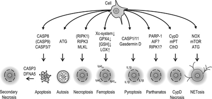
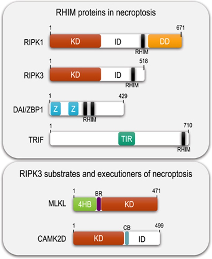
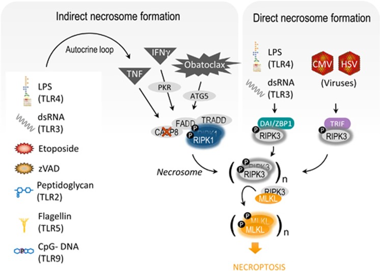
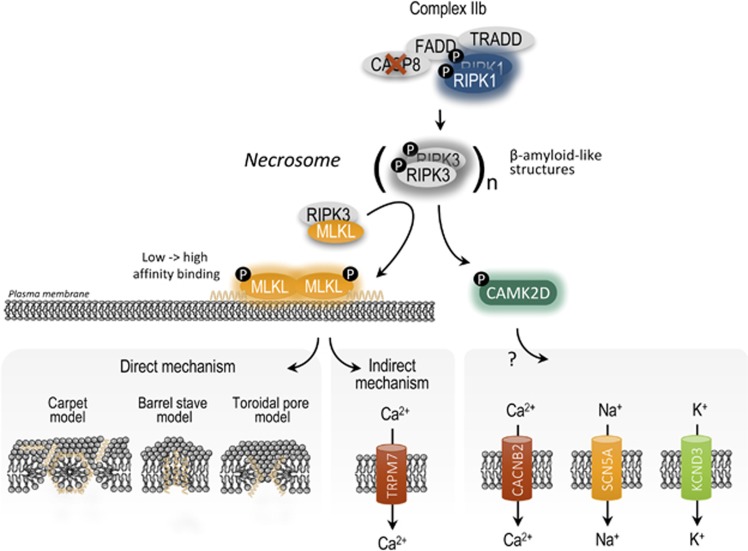
Necroptosis is a form of regulated cell death, which is induced by ligand binding to TNF family death domain receptors, pattern recognizing receptors and virus sensors. The common feature of these receptor systems is the implication of proteins, which contain a receptor interaction protein kinase (RIPK) homology interaction motif (RHIM) mediating recruitment and activation of receptor-interacting protein kinase 3 (RIPK3), which ultimately activates the necroptosis executioner mixed lineage kinase domain-like (MLKL). In case of the TNF family members, the initiator is the survival- and cell death-regulating RIPK1 kinase, in the case of Toll-like receptor 3/4 (TLR3/4), a RHIM-containing adaptor, called TRIF, while in the case of Z-DNA-binding protein ZBP1/DAI, the cytosolic viral sensor itself contains a RHIM domain. In this review, we discuss the different protein complexes that serve as nucleation platforms for necroptosis and the mechanism of execution of necroptosis. Transgenic models (knockout, kinase-dead knock-in) and pharmacologic inhibition indicate that RIPK1, RIPK3 or MLKL are implicated in many inflammatory, degenerative and infectious diseases. However, the conclusion of necroptosis being solely involved in the etiology of diseases is blurred by the pleiotropic roles of RIPK1 and RIPK3 in other cellular processes such as apoptosis and inflammasome activation.


Conflict of interest statement

The authors declare no conflict of interest.

Figures

Figure 1
Figure 1
An overview of regulated cell death modalities and the core pathway proteins involved. Depending on cell type and intra/extracellular conditions, cells can activate different cell death modalities with different core pathway proteins involved. Cell death modalities with an apoptotic or an autophagic morphology are considered to be less immunogenic than cell death with a necrotic morphology, because the former two are consisting of containment programs aimed at preventing the release of intracellular content and associated with efficient phagocytosis, while necrotic cell death is essentially associated with membrane permeabilization resulting the rapid release of the cellular contents in the environment acting as danger or damage-associated molecular patterns (represented by a gray halo) and attracting immune cells. Note that in many conditions, induction of autophagy is not associated with cell death, but rather a homeostasis mechanism following cellular stress. However, in some conditions, autophagy may proceed to cell death and targeting Atg genes will retard in such cases the cell death process. AIF, apoptosis-inducing factor; ATG, autophagy related; CASP, caspase; CypD, cyclophillin D; DFNA5, deafness-associated tumor suppressor; GPX4, glutathion peroxidase 4; GSH, reduced glutathion; IL1β, interleukin-1 β; LOX, lipoxygenase; lipidox, peroxidized (phospho)lipids; MLKL, mixed lineage kinase domain-like; mPT, mitochondrial permeability transition pore; mTOR, mechanistic target of rapamycin; NOX, NADPH oxidase; PARP, poly-(ADP ribose)-polymerase; RIPK, receptor-interacting protein kinase; Xc-system, cystin/glutamate antiporter
Figure 2
Figure 2
Domain structure of the key mediators of necroptosis. Length is indicated in number of amino acids. 4HB, four-helical bundle domain; BR, brace region; CAMK2D, calcium/calmodulin-dependent protein kinase type II subunit delta; CB, calmodulin-binding domain; DD, death domain; ID, intermediate domain; KD, kinase domain; MLKL, mixed lineage kinase domain-like; RHIM, RIP homotypic interaction motif; RIPK, receptor-interacting protein kinase; TIR, Toll/interleukin receptor domain; TRIF, TIR domain-containing adaptor-inducing IFN-beta (also known as TICAM1, TIR domain-containing adapter molecule 1); ZBP1, Z-DNA-binding protein 1 (also known as DAI, DNA-dependent activator of IFN-regulatory factors)
Figure 3
Figure 3
Signal transduction complexes in TNF-induced survival and cell death. (a) TNF stimulation results in formation of a receptor bound complex I, which essentially prevents cell death (see text). (b) TNF receptor dissociation of RIPK1 results in the formation of different pro-cell death complexes (complex IIa, IIb and the necrosome). Complex IIa contains TRADD and can be formed independent of the scaffold and kinase function of RIPK1. In contrast, complex IIb lacks TRADD and requires kinase-active RIPK1 for cell death induction. It is still unclear whether these complexes represent different physical entities or different compositions or post-translational regulations of essentially the same complex II. CASP, caspase; cIAP, cellular inhibitor of apoptosis protein; FADD, Fas-associated protein with death domain; HOIL-1 or RBCK1, RANBP2-type and C3HC4-type zinc-finger-containing 1; HOIP or RNF31, ring finger protein 31; IKK, inhibitor of NF-κB kinase; MLKL, mixed lineage kinase domain-like; NEMO, NF-κB essential modulator (sometimes misleadingly called IKKγ since it does not possess kinase activity); NF-κB, nuclear factor kappa B; Otulin, OTU deubiquitinase with linear linkage specificity; PELI1, pellino 1; RIPK, receptor-interacting protein kinase; Sharpin, SHANK-associated RH domain-interacting protein; TAB, TAK1-binding protein; TAK1, TGFβ activated kinase 1; TNF, tumor necrosis factor; TNFR, TNF receptor; TRADD, TNF-associated death domain
Figure 4
Figure 4
Direct and indirect necrosome formation by different stimuli. Many stimuli elicit necroptosis in a direct and indirect way. The direct necrosome formation involves the canonical pathway that requires RIPK1 kinase activity and the non-canonical pathway that is dependent on the TRIF adaptor or the ZBP1/DAI sensor. In the indirect way, stimuli elicit the production of TNF, which on its turn activates the canonical pathway. ATG5, autophagy related; CASP8, caspase; CMV, cytomegalovirus; FADD, Fas-associated protein with death domain; HSV, human Simian virus; IFNγ, interferon gamma; LPS, lipopolysaccharide; MLKL, mixed lineage kinase domain-like; PKR, dsRNA-activated protein kinase R (officially known as EIF2AK2); RIPK, receptor-interacting protein kinase; TLR, Toll-like receptor; TNF, tumor necrosis factor; TRADD, TNF-associated death domain; TRIF, officially known as Toll-like receptor adaptor molecule (TICAM); ZBP1, Z-DNA-binding protein 1 (also known as DAI); zVAD, pan-caspase inhibitor
Figure 5
Figure 5
Necrosome formation and execution of necroptosis. RHIM-dependent necrosome formation results in phosphorylation and activation of MLKL or CAMK2 by RIPK3, which in turn activate ion influxes in the cell. MLKL is initially recruited to the plasma membrane by weak interactions with phospholipids (PIPs), but after a conformational change a second, stronger PIP interaction site is revealed in MLKL. MLKL can either directly form pores in the plasma membrane or activate ion channels, whereas CAMK2D is a known activator of several ion channels (without a currently demonstrated role in necroptosis). How MLKL induces permeabilization of the plasma membrane is unknown, but may be similar to other types of pore-forming proteins (carpet, barrel or toroidal model). 4HBD, four-helical bundle domain; CAC4NB, L-type Ca2+ channel subunit beta-2; CAMK2D, calcium-dependent protein kinase II delta; CASP, caspase; FADD, Fas-associated protein with death domain; KCND3, K+ channel potassium channel, voltage gated Shal-related subfamily D; MLKL, mixed lineage kinase domain-like; RHIM, RIPK homotypic interaction motif; RIPK, receptor-interacting protein kinase; SCN5A, sarcolemmal cardiac Na+ channel; TRADD, TNF-associated death domain; TRPM7, transient receptor potential cation channel, subfamily M, member 7; ZBP1, Z-DNA-binding protein 1 (also known as DAI)

References

    1. Vaux DL, Korsmeyer SJ. Cell death in development. Cell 1999; 96: 245–254. - PubMed
    1. Glucksmann A. Cell deaths in normal vertebrate ontogeny. Biol Rev Camb Philos Soc 1951; 26: 59–86. - PubMed
    1. Renehan AG, Booth C, Potten CS. What is apoptosis, and why is it important? BMJ 2001; 322: 1536–1538. - PMC - PubMed
    1. Hanahan D, Weinberg RA. Hallmarks of cancer: the next generation. Cell 2011; 144: 646–674. - PubMed
    1. Manji GA, Hozak RR, LaCount DJ, Friesen PD. Baculovirus inhibitor of apoptosis functions at or upstream of the apoptotic suppressor P35 to prevent programmed cell death. J Virol 1997; 71: 4509–4516. - PMC - PubMed

Substances

LinkOut - more resources