. 2017 Jul;16(1):174-180.
doi: 10.3892/mmr.2017.6580. Epub 2017 May 12.

Mitochondrial activity and oxidative stress functions are influenced by the activation of AhR-induced CYP1A1 overexpression in cardiomyocytes

Affiliations

Mitochondrial activity and oxidative stress functions are influenced by the activation of AhR-induced CYP1A1 overexpression in cardiomyocytes

Bing Zhou et al. Mol Med Rep. 2017 Jul.

Abstract

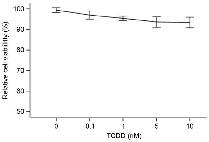
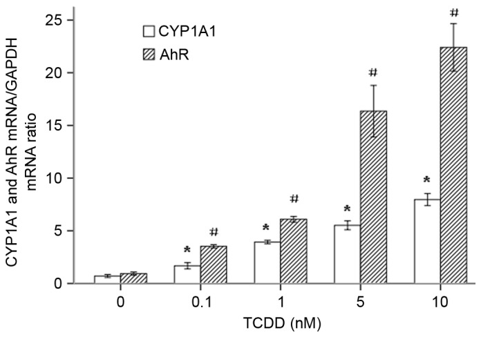
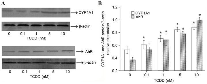
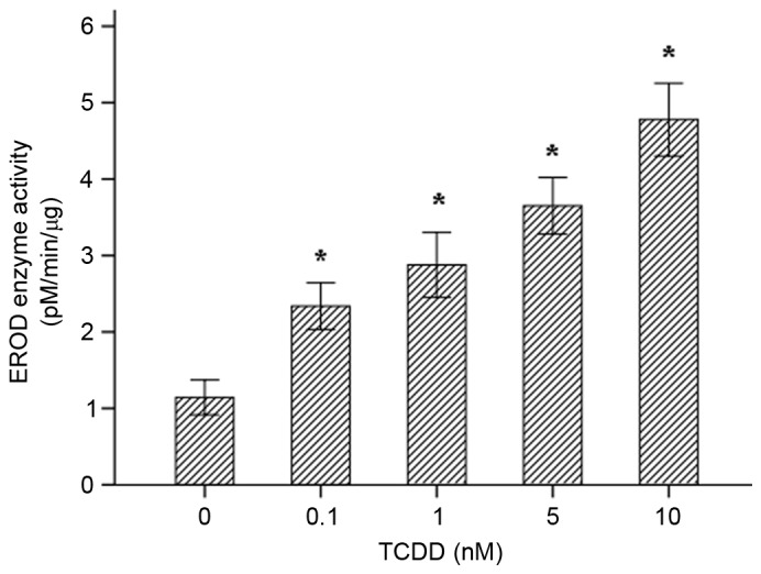
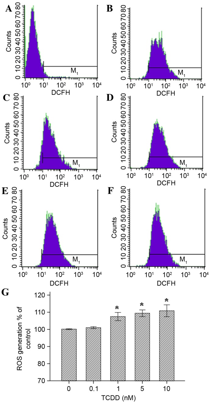
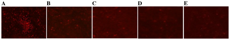
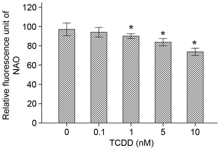
There is an endemic cardiomyopathy currently occurring in China, termed, Keshan disease (KD). The authors previously compared mitochondrial‑associated gene expression profiles of peripheral blood mononuclear cells (PBMCs) derived from KD patients and normal controls, using mitochondria‑focused cDNA microarray technology. The results detected an upregulation of the enzyme‑associated CYP1A1 gene, (ratios ≥2.0). The aryl hydrocarbon receptor (AhR) regulates the expression of numerous cytochrome P450 (CYP) genes including members of the CYP1 family; CYP1A1 and CYP1A2. Several previous studies have suggested roles for the aryl hydrocarbon receptor (AhR) and the genes that it regulates. An example involves cytochrome P4501A1 (CYP1A1), in the pathogenesis of heart failure, cardiac hypertrophy and other cardiomyopathies. Mitochondria comprise ~30% of the intracellular volume in mammalian cardiomyocytes, and subtle alterations in mitochondria can markedly influence cardiomyopathies. The present study investigated alterations in the activity and functions of mitochondria following AhR‑induced overexpression of CYP1A1. AC16 cells were treated with the CYP1A1 inducer 2,3,7,8‑tetrachlorodibenzo‑p‑dioxin (TCDD), and cytotoxicity was then evaluated in MTT assays. Reverse transcription‑quantitative polymerase chain reactions, western blot analysis and 7‑ethoxyresorufin O‑deacylase assays were performed to analyze the mRNA and protein levels, and the enzymatic activity of CYP1A1. Mitochondrial activity and mass were analyzed using an inverted fluorescence microscope and a fluorescence microplate reader. Reactive oxygen species (ROS) activity was analyzed using flow cytometry. The results of the current study demonstrated that TCDD gradually increased mRNA and protein levels of AhR and CYP1A1, in addition to the enzymatic activity. Mitochondrial activity and the quality of mitochondrial membranes were also significantly attenuated, and mitochondrial ROS levels were elevated in the TCDD‑induced cardiomyocytes. The results indicate the involvement of the AhR/CYP1A1 signaling pathway in the mechanism of action of TCDD in human cardiomyocytes. The present findings may provide an explanation for myocardial injuries caused by polycyclic aromatic hydrocarbons. The authors conclude that exposure to TCDD results in regulatory alteration to the expression of detoxification genes that ultimately affect the metabolic activation and function of cardiomyocytes.


Figures

Figure 1.
Figure 1.
Effects of TCDD on cell viability. Cells were treated with various concentrations of TCDD (0, 0.1, 1, 5 and 10 nM) for 24 h and then analyzed using MTT assays. Treating cells with TCDD produced no adverse effects on cell viability when the treated cells were compared with control cells that were incubated with only culture medium (100% viability). P>0.05 vs. control. TCDD, 2,3,7,8-tetrachlorodibenzo-p-dioxin.
Figure 2.
Figure 2.
Relative gene expression ratios for the CYP1A1 and AhR mRNAs in TCDD-treated AC16 cardiomyocytes. The cells were pre-treated with various concentrations of TCDD for 24 h. P<0.05 for the expression level of CYP1A1 in each treatment group when compared with the cells that were incubated with only culture medium (*P<0.05 vs. untreated cells); #P<0.05 for the expression level of AhR in each treatment group when compared with the cells that were incubated with culture medium alone (#P<0.05 vs. untreated cells). ArH, aryl hydrocarbon receptor; TCDD, 2,3,7,8-tetrachlorodibenzo-p-dioxin.
Figure 3.
Figure 3.
Effects of TCDD on the protein expression of CYP1A1 and AhR in AC16 cardiomyocytes. Cells were incubated for 24 h with various concentrations of TCDD (0, 0.1, 1, 5 and 10 nM). (A) The proteins (25 µg) were separated using 10% SDS-PAGE. The protein levels of CYP1A1 and AhR were determined using western blot analysis. β-actin was used as the loading control. (B) The bar graph represents the relative normalized levels of the CYP1A1 and AhR proteins. The results are expressed as a percentage of the control values, which were determined to represent 100%. P<0.05 for the expression of CYP1A1 in each treatment group compared to the expression observed in the untreated cells (*P<0.05 vs. untreated cells). #P<0.05 for the expression of AhR in each treatment group compared with the expression observed in the cells that were incubated with only culture medium (#P<0.05 vs. untreated cells). TCDD, 2,3,7,8-tetrachlorodibenzo-p-dioxin; ArH, aryl hydrocarbon receptor.
Figure 4.
Figure 4.
CYP1A1 activity was measured in intact living cells using 96-well cell culture plates and 7-ethoxyresorufin as the substrate. The effects of TCDD treatment on the EROD activity of CYP1A1 in AC16 cells (mean ± standard deviation, n=3) are presented. *P<0.05 vs. control group. TCDD, 2,3,7,8-tetrachlorodibenzo-p-dioxin; EROD, 7-ethoxyresorufin O-deacylase.
Figure 5.
Figure 5.
Effects of TCDD on the generation of ROS in AC16 cardiomyocytes that were pre-treated with TCDD for 24 h. Intracellular ROS levels were estimated using flow cytometry with the probe DCFH-DA, which is oxidized to form DCF in the presence of ROS. The cells were treated as follows: (A) Blank group (non-probe); (B) control (concentration=0 nM); (C) intervention group, (0.1 nM); (D) intervention groups (1 nM); (E intervention group (5 nM); and (F) intervention group (10 nM). (G) A guidance line has been drawn through the histograms being compared. TCDD, 2,3,7,8-tetrachlorodibenzo-p-dioxin; ROS, reactive oxygen species; DCFH-DA, 2′,7′-dichlorofluorescein diacetate; DCF, dichlorofluorescein.
Figure 6.
Figure 6.
Effects of TCDD on mitochondrial activity in AC16 cells. The mitochondria of living cells were stained using Mito Tracker RED. Digital images of each well were obtained using inverted fluorescence microscopy at ×40 magnification. (A), (B), (C), (D) and (E) represent 0, 0.1, 1, 5 and 10 nM of TCDD, respectively. TCDD, 2,3,7,8-tetrachlorodibenzo-p-dioxin.
Figure 7.
Figure 7.
Mitochondria mass was measured in living cells using 10-N-nonyl acridine orange as a substrate. Fluorescence intensities were assessed using a microplate reader. *P<0.05 vs. Control group (concentration=0 nM). TCDD, 2,3,7,8-tetrachlorodibenzo-p-dioxin.

References

    1. Korashy HM, El-Kadi AO. The role of aryl hydrocarbon receptor in the pathogenesis of cardiovascular diseases. Drug Metab Rev. 2006;38:411–450. doi: 10.1080/03602530600632063. - DOI - PubMed
    1. Kann S, Huang MY, Estes C, Reichard JF, Sartor MA, Xia Y, Puga A. Arsenite-induced aryl hydrocarbon receptor nuclear translocation results in additive induction of phase I genes and synergistic induction of phase II genes. Mol Pharmacol. 2005;68:336–346. - PubMed
    1. Nebert DW, Dalton TP, Okey AB, Gonzalez FJ. Role of aryl hydrocarbon receptor-mediated induction of the CYP1 enzymes in environmental toxicity and cancer. J Biol Chem. 2004;279:23847–23850. doi: 10.1074/jbc.R400004200. - DOI - PubMed
    1. Fujii-Kuriyama Y, Mimura J. Molecular mechanisms of AhR functions in the regulation of cytochrome P450 genes. Biochem Biophys Res Commun. 2005;338:311–317. doi: 10.1016/j.bbrc.2005.08.162. - DOI - PubMed
    1. Thum T, Borlak J. Testosterone, cytochrome P450 and cardiac hypertrophy. FASEB J. 2002;16:1537–1549. doi: 10.1096/fj.02-0138com. - DOI - PubMed

MeSH terms

Substances