. 2017 Nov 17;8(8):1668-1682.
doi: 10.1080/21505594.2017.1330240. Epub 2017 Jun 2.

The distinct function of Tep2 and Tep6 in the immune defense of Drosophila melanogaster against the pathogen Photorhabdus

Affiliations

The distinct function of Tep2 and Tep6 in the immune defense of Drosophila melanogaster against the pathogen Photorhabdus

Upasana Shokal et al. Virulence. .

Abstract

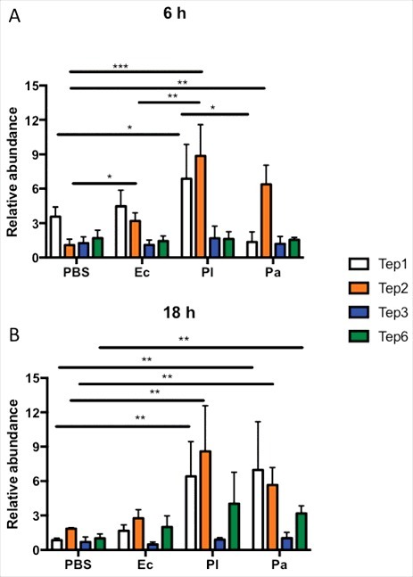
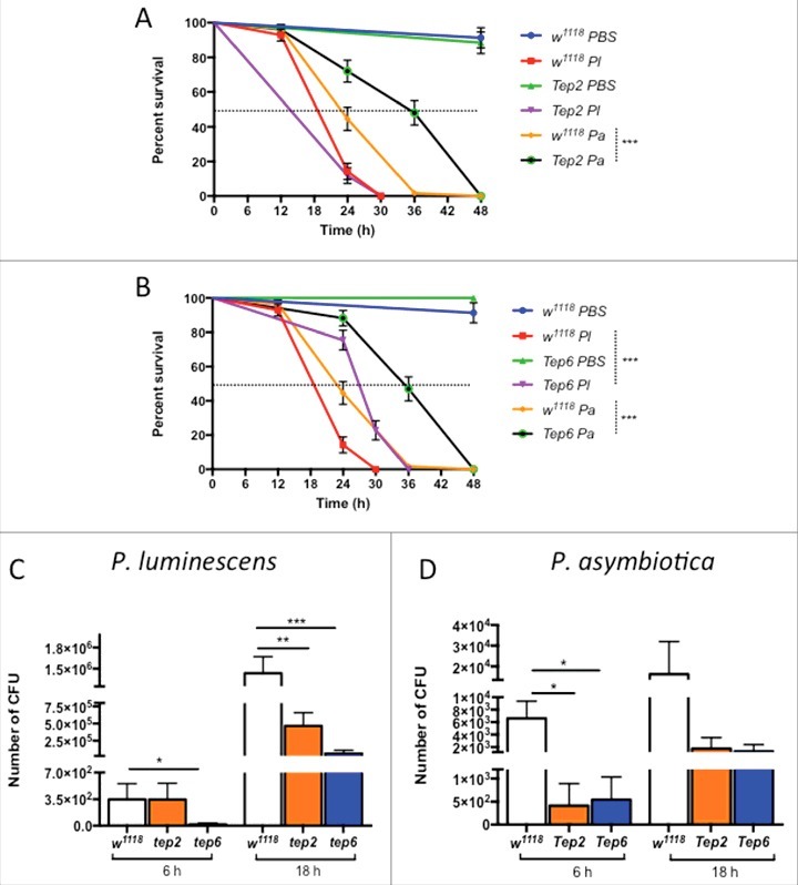
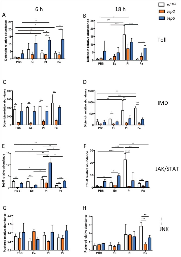
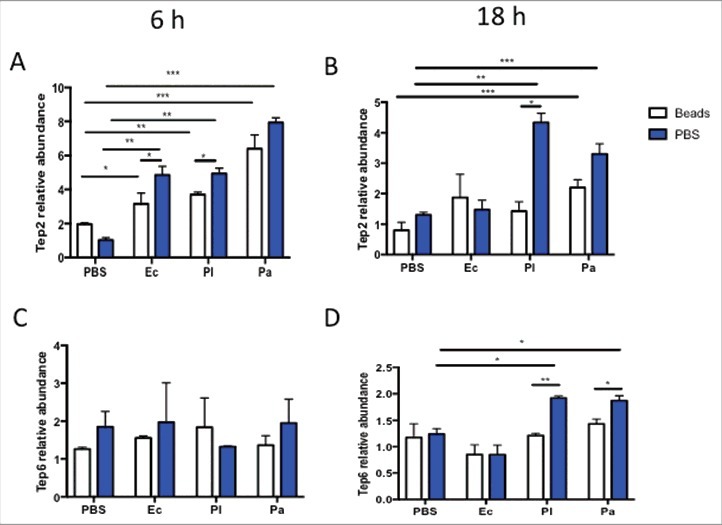
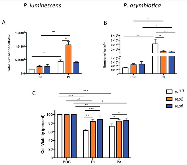
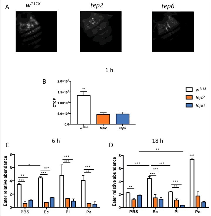
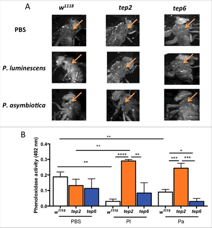
Previous and recent investigations on the innate immune response of Drosophila have identified certain mechanisms that promote pathogen elimination. However, the function of Thioester-containing proteins (TEPs) in the fly still remains elusive. Recently we have shown the contribution of TEP4 in the antibacterial immune defense of Drosophila against non-pathogenic E. coli, and the pathogens Photorhabdus luminescens and P. asymbiotica. Here we studied the function of Tep genes in both humoral and cellular immunity upon E. coli and Photorhabdus infection. We found that while Tep2 is induced after Photorhabdus and E. coli infection; Tep6 is induced by P. asymbiotica only. Moreover, functional ablation of hemocytes results in significantly low transcript levels of Tep2 and Tep6 in response to Photorhabdus. We show that Tep2 and Tep6 loss-of-function mutants have prolonged survival against P. asymbiotica, Tep6 mutants survive better the infection of P. luminescens, and both tep mutants are resistant to E. coli and Photorhabdus. We also find a distinct pattern of immune signaling pathway induction in E. coli or Photorhabdus infected Tep2 and Tep6 mutants. We further show that Tep2 and Tep6 participate in the activation of hemocytes in Drosophila responding to Photorhabdus. Finally, inactivation of Tep2 or Tep6 affects phagocytosis and melanization in flies infected with Photorhabdus. Our results indicate that distinct Tep genes might be involved in different yet crucial functions in the Drosophila antibacterial immune response.

Keywords: Drosophila; Photorhabdus; immunity; thioester-containing protein.


Figures

Figure 1.
Figure 1.
Tep1, Tep2 and Tep6 genes are upregulated in D. melanogaster flies by Photorhabdus infection. Transcript levels of Tep1, Tep2, Tep3 and Tep6 genes are shown in w1118 flies (n = 3–5) after (A) 6 and (B) 18 hpi with 1XPBS (septic injury control), E. coli (Ec), P. luminescens (Pl) and P. asymbiotica (Pa). Gene transcript levels are shown as relative abundance of transcripts normalized to RpL32 and expressed as a ratio compared with untreated flies (negative control). Significant differences are shown with asterisks (*p < 0.05, **p < 0.01, ***p < 0.001). Bars show the means from 3 independent experiments and error bars represent standard deviation.
Figure 2.
Figure 2.
Survival and bacterial load analysis for tep2 and tep6 mutants after Photorhabdus infection. Survival curves for loss-of-function (A) tep2 mutants and (B) tep6 mutants with w1118 (background control flies) are shown. Flies (n = 20) were injected in the thorax by microinjection with 1XPBS (septic injury control), P. luminescens (Pl) or P. asymbiotica (Pa). Survival was monitored at 6 h intervals for 48 h. The black dotted line represents 50% survival. Colony forming units (CFU) of (C) P. luminescens and (D) P. asymbiotica are shown in tep2, tep6 and control flies (n = 5 per experimental condition) after 6 and 18 hpi. CFU were quantified through quantitative PCR of makes caterpillars floppy (mcf-1) in P. luminescens and the insecticidal toxin complex protein gene (tccC3) in P. asymbiotica. Significant differences are indicated with asterisks (* p < 0.05, ** p <0.01, *** p <0.001). The means from 3 independent experiments are shown and error bars represent standard errors (survival) and standard deviation (bacterial load).
Figure 3.
Figure 3.
D. melanogaster Tep2 and Tep6 differentially regulate the activation of immune pathways against Photorhabdus. Transcript levels for (A, B) Defensin (Toll pathway), (C, D) Diptericin (IMD pathway), (E, F) Tot-M (JAK/STAT pathway) and (G, H) Puckered (JNK pathway) in loss-of function tep2 and tep6 mutants with their corresponding control strain (w1118) at 6 and 18 hpi with 1XPBS, E. coli (Ec), P. luminescens (Pl) or P. asymbiotica (Pa) (n = 3 individuals per experimental condition). Gene transcript levels are shown as relative abundance of transcripts normalized to RpL32 and expressed as a ratio compared with untreated flies (negative control). Values represent the means from 3 biologic replicates and error bars represent standard deviations. Significant differences are indicated with asterisks; *p <0.05, **p <0.01, ***p <0.001, ****p <0.0001).
Figure 4.
Figure 4.
Transcript levels of Tep2 and Tep6 are significantly decreased in control flies (w1118) with functionally ablated hemocytes. Transcript levels of (A) Tep2 and (B) Tep6 at 6 and 18 hpi with 1XPBS, E. coli (Ec), P. luminescens (Pl) or P. asymbiotica (Pa) in w1118 flies (n = 5) pre-injected with beads or 1XPBS. Significant differences are indicated with asterisks (*p < 0.05, ** p < 0.01, ***p < 0.001). Bars show the means from 2 independent experiments and error bars represent standard deviations.
Figure 5.
Figure 5.
D. melanogaster tep2 and tep6 mutants display variable number of total hemocyte counts and increased hemocyte viability compared with control flies (w1118) after Photorhabdus infection. According to the hemocyte counting protocol, total number of hemocytes (total cells/ml) in tep2 and tep6 mutants with control flies after 18 h of injection with 1XPBS, (A) P. luminescens (Pl) or (B) P. asymbiotica (Pa). The percentage of total viable cells in the control and tep mutant flies at 18 hpi with 1XPBS, (C) P. luminescens and (D) P. asymbiotica. Significant differences are indicated with asterisks (*p < 0.05, **p < 0.01, ***p < 0.001). Bars show the means from 3 independent experiments and error bars represent standard deviations.
Figure 6.
Figure 6.
Tep2 and Tep6 are essential for the phagocytosis process in Drosophila. (A) Representative images of phagocytosis in tep2 and tep6 loss-of-function mutants and control flies (w1118) at 1 hpi of lipophilized pHrodo-labeled E. coli particles. Images were taken using fluorescence microscopy at 10X magnification. (B) Corrected total cell fluorescence (CTCF) in tep mutants and w1118 flies (n = 7), 1 h following injection of pHrodo-labeled E. coli. Images were processed in ImageJ and CTCF was estimated. Transcript levels of Eater in tep mutants and w1118 flies (n = 5) at (C) 6 h and (D) 18 hpi of 1XPBS, E. coli (Ec), P. luminescens (Pl) or P. asymbiotica (Pa). Significant differences are indicated with asterisks (*p < 0.05, **p < 0.01, ***p < 0.001). The means from 2 (Eater transcription) -three (Phagocytosis) independent experiments are shown and error bars represent standard deviation.
Figure 7.
Figure 7.
Melanization response and PO activity are elevated in D. melanogaster tep2 mutants upon Photorhabdus infection. (A) Melanization of the wound site in tep2 and tep6 loss-of-function mutant flies and their background control strains (w1118) is shown at 10X magnification 3 h after injection with PBS, P. luminescens or P. asymbiotica bacteria. Arrows indicate the site of injury. (B) PO activity in the hemolymph plasma of tep2, tep6 mutants and control flies (w1118) at 3 hpi with PBS, P. luminescens (Pl) or P. asymbiotica (Pa) (n = 20 flies) as measured by the optical density at 492 nm after incubation with L-Dopa. Values represent the means from 3 biologic replicates and error bars represent standard deviations. Significant differences are indicated with asterisks (*p < 0.05, **p < 0.01, ***p < 0.001, ****p < 0.0001). The means from 3 independent experiments are shown and error bars represent standard deviation.

Comment in

Similar articles

Cited by

References

    1. Lemaitre B, Hoffmann J. The host defense of Drosophila melanogaster. Ann Rev Immunol 2007; 25:697-743; PMID:17201680; https://doi.org/ 10.1146/annurev.immunol.25.022106.14161510.1146/annurev.immunol.25.022106.141615 - DOI - PubMed
    1. Pal S, Wu LP. Pattern recognition receptors in the fly: lessons we can learn from the Drosophila melanogaster immune system. Fly (Austin) 2009; 3:121-9; PMID:19440043; https://doi.org/ 10.4161/fly.882710.4161/fly.8827 - DOI - PubMed
    1. Buresova V, Hajdusek O, Franta Z, Sojka D, Kopacek P. IrAM-An alpha2-macroglobulin from the hard tick Ixodes ricinus: characterization and function in phagocytosis of a potential pathogen Chryseobacterium indologenes. Dev Comp Immunol 2009; 33:489-98; PMID:18948134; https://doi.org/ 10.1016/j.dci.2008.09.01110.1016/j.dci.2008.09.011 - DOI - PubMed
    1. Blandin S, Shiao SH, Moita LF, Janse CJ, Waters AP, Kafatos FC, Levashina EA. Complement-like protein TEP1 is a determinant of vectorial capacity in the malaria vector Anopheles gambiae. Cell 2004; 116:661-70; PMID:15006349; https://doi.org/ 10.1016/S0092-8674(04)00173-410.1016/S0092-8674(04)00173-4 - DOI - PubMed
    1. Xiao X, Liu Y, Zhang X, Wang J, Li Z, Pang X, Wang P, Cheng G. Complement-related proteins control the flavivirus infection of Aedes aegypti by inducing antimicrobial peptides. PLoS Pathog 2014; 10:e1004027; PMID:24722701; https://doi.org/ 10.1371/journal.ppat.100402710.1371/journal.ppat.1004027 - DOI - PMC - PubMed

Publication types

LinkOut - more resources