. 2017 May 12;12(5):e0176280.
doi: 10.1371/journal.pone.0176280. eCollection 2017.

Assessment of the quality of DNA from various formalin-fixed paraffin-embedded (FFPE) tissues and the use of this DNA for next-generation sequencing (NGS) with no artifactual mutation

Affiliations

Assessment of the quality of DNA from various formalin-fixed paraffin-embedded (FFPE) tissues and the use of this DNA for next-generation sequencing (NGS) with no artifactual mutation

Naoki Einaga et al. PLoS One. .

Abstract

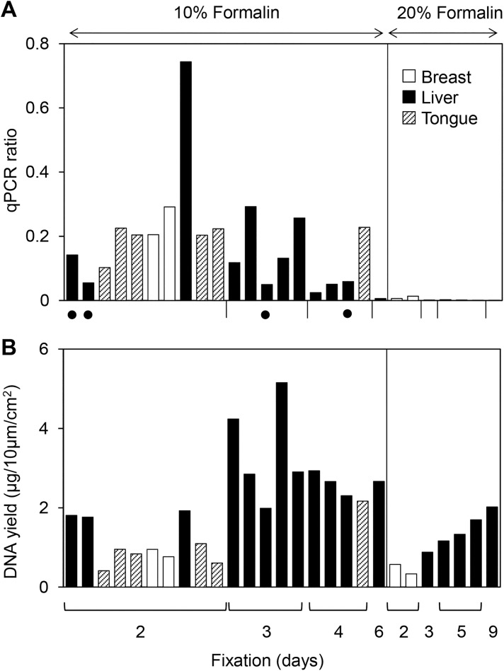
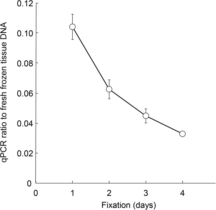
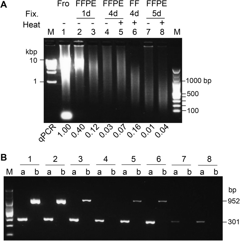
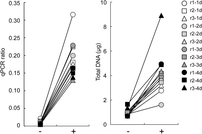
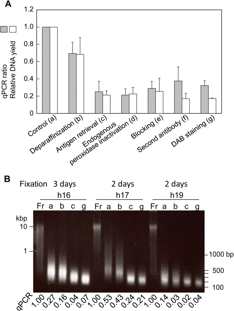
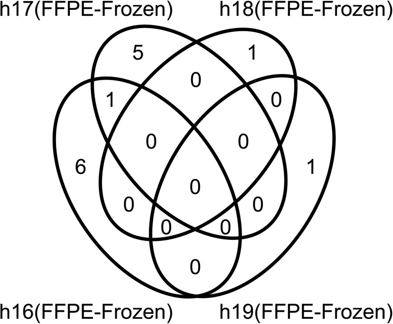
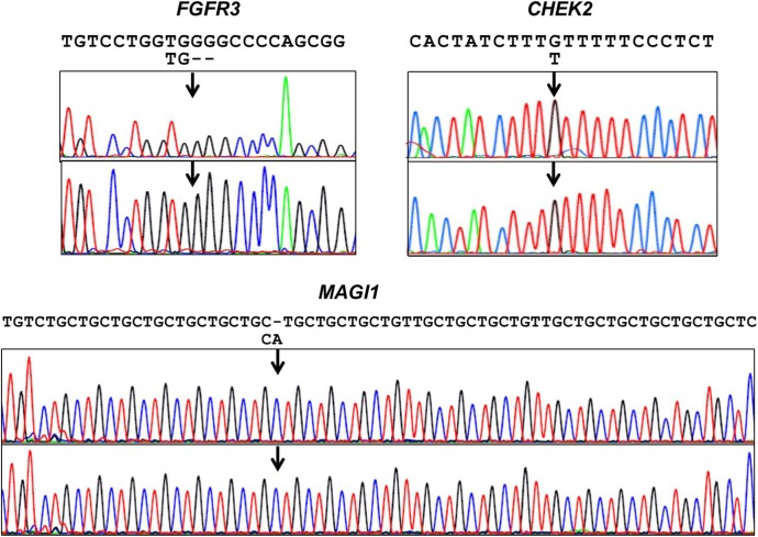
Formalin-fixed, paraffin-embedded (FFPE) tissues used for pathological diagnosis are valuable for studying cancer genomics. In particular, laser-capture microdissection of target cells determined by histopathology combined with FFPE tissue section immunohistochemistry (IHC) enables precise analysis by next-generation sequencing (NGS) of the genetic events occurring in cancer. The result is a new strategy for a pathological tool for cancer diagnosis: 'microgenomics'. To more conveniently and precisely perform microgenomics, we revealed by systematic analysis the following three details regarding FFPE DNA compared with paired frozen tissue DNA. 1) The best quality of FFPE DNA is obtained by tissue fixation with 10% neutral buffered formalin for 1 day and heat treatment of tissue lysates at 95°C for 30 minutes. 2) IHC staining of FFPE tissues decreases the quantity and quality of FFPE DNA to one-fourth, and antigen retrieval (at 120°C for 15 minutes, pH 6.0) is the major reason for this decrease. 3) FFPE DNA prepared as described herein is sufficient for NGS. For non-mutated tissue specimens, no artifactual mutation occurs during FFPE preparation, as shown by precise comparison of NGS of FFPE DNA and paired frozen tissue DNA followed by validation. These results demonstrate that even FFPE tissues used for routine clinical diagnosis can be utilized to obtain reliable NGS data if appropriate conditions of fixation and validation are applied.


Conflict of interest statement

Competing Interests: The authors have declared that no competing interests exist.

Figures

Fig 1
Fig 1. DNA quality and quantity of various clinical diagnostic FFPE tissues.
DNA was extracted from 27 FFPE samples of normal breast, normal liver and tumorous tongue and then qualified and quantified. The samples were divided into two groups to test the formalin concentration and subdivided for the number of fixation days. Six tongue tissues were fixed with 10% neutral buffered formalin. The four samples with black dots were fixed at a temperature higher than 25°C. (A) The DNA quality was determined by qPCR of human GAPDH (93 bp), and the results are expressed as the ratio to fresh-frozen DNA. (B) The DNA yield was determined using an UV spectrophotometer, and the results are expressed as DNA quantity per cm2 of tissue area with 10 μm thickness.
Fig 2
Fig 2. FFPE DNA quality is dependent on the formalin fixation duration.
Three sets of 5 matched samples containing fresh-frozen tissue DNA and 4 FFPE (1-, 2-, 3- and 4-day fixation) DNA samples were prepared from 3 rat liver specimens (r1, r2, r3) and examined for DNA quality. The data are shown as averages (circles) with standard deviations (bars).
Fig 3
Fig 3. Agarose gel electrophoresis of rat liver DNA.
(A) Agarose gel electrophoresis (0.8%) of 400 ng of purified DNA. DNA on the gel were stained with GelRed which is sensitive to double-stranded DNA. The qPCR ratio to matched frozen tissue DNA is shown at the bottom of each lane. Fro, frozen tissue DNA; Fix., fixation for 1 day (1 d): lanes 2 and 3 were different tissue samples with 1 day fixation, 4 days (4 d) and 5 days (5 d); Heat, heat treatment of lysate (+) or not (-) at 95°C for 30 minutes; M, molecular weight marker; qPCR, qPCR ratio to frozen tissue DNA. (B) Agarose gel electrophoresis (2%) of PCR products with target sizes of 301 bp (a) and 952 bp (b). The rat Tp53 gene was amplified from the indicated DNA, lanes 1 to 8 in (A).
Fig 4
Fig 4. Effects of heat treatment of FFPE lysates on DNA quality and quantity.
(A) Rat liver FFPE specimens: 6 matched frozen and FFPE tissues (r1 to r3 fixed for 4 days and r4 to r6 fixed for 5 days shown in S1 Fig) were used, and the FFPE tissue lysates were treated (+) or not (-) at 95°C for 30 minutes before purification. DNA quality and quantity were determined as shown in Fig 1. (B) Human normal liver FFPE specimens: 4 matched frozen and FFPE tissues (h16, h18, h19 and h22) were used, and the FFPE tissue lysates were heat treated and examined for DNA quality (left) and quantity (right), as described in (A). **, p<0.01; *, p<0.05 by the paired t-test.
Fig 5
Fig 5. FF DNA quality and quantity dependent on the heat treatment.
Three sets of 5 matched samples containing fresh-frozen and FF (1-, 2-, 3- and 4-day fixation) tissues were prepared from 3 rat liver specimens (r1, r2, r3). FF DNA was extracted from the lysates with (+) or without (-) heat treatment and examined for DNA quality (left) and DNA quantity (right).
Fig 6
Fig 6. DNA quality and quantity during the course of IHC staining of FFPE thin sections.
(A) The DNA qPCR ratio and relative DNA yield were expressed using FFPE thin sections before the start of IHC used as the control. The IHC procedure was divided into 6 steps. Four FFPE samples (h16 to h19) were used for human hepatocyte marker immunostaining. The qPCR value and DNA yield relative to the matched control were calculated, and the mean values of 4 samples were compared among the 6 steps. Control DNA was prepared directly from matched FFPE thin sections according to our modified RecoverAll protocol with additional heat treatment. (B) Agarose gel (0.8%) electrophoresis of 3 representative sets (h16, h17 and h19) of matched DNA samples (500 ng/lane). Fr, frozen tissue DNA; a, b, c, g, DNAs as shown in (A).
Fig 7
Fig 7. Venn diagram of variant candidates found in 4-pair analyses of FFPE and frozen tissue DNA.
Variants identified in 4 matched FFPE-frozen pairs of normal liver tissues, h16, h17, h18 and h19. Fourteen variants were identified by pair analysis using Ion Reporter version 5.0 with the following filters: coverage more than 100, allele frequency more than 0.05, frozen allele coverage = 0. ERCC2 was a common variant among two samples, h16 and h17.
Fig 8
Fig 8. Sanger sequencing of FGFR3, CHEK2, and MAGI1.
Variant calls at high frequency were invalidated by Sanger sequencing of amplified products from matched fresh-frozen DNA (upper panel) and FFPE DNA (lower panel). Arrows indicate the positions of a deletion (FGFR3), an SNV (CHEK2) and an insertion (MAGI1), and the variant sequences are shown below the reference sequences.

Similar articles

Cited by

References

    1. Vogelstein B, Papadopoulos N, Velculescu VE, Zhou S, Diaz LA Jr., Kinzler KW. Cancer genome landscapes. Science. 2013; 339(6127): 1546–58. PubMed Central PMCID: PMC3749880. doi: 10.1126/science.1235122 - DOI - PMC - PubMed
    1. Garraway LA, Lander ES. Lessons from the cancer genome. Cell. 2013; 153(1): 17–37. doi: 10.1016/j.cell.2013.03.002 - DOI - PubMed
    1. Do H, Dobrovic A. Sequence artifacts in DNA from formalin-fixed tissues: causes and strategies for minimization. Clin Chem. 2015; 61(1): 64–71. doi: 10.1373/clinchem.2014.223040 - DOI - PubMed
    1. Nakayama Y, Yamaguchi H, Einaga N, Esumi M. Pitfalls of DNA Quantification Using DNA-Binding Fluorescent Dyes and Suggested Solutions. PLoS One. 2016; 11(3): e0150528 PubMed Central PMCID: PMC4777359. doi: 10.1371/journal.pone.0150528 - DOI - PMC - PubMed
    1. Wong SQ, Li J, Tan AY, Vedururu R, Pang JM, Do H, et al. Sequence artefacts in a prospective series of formalin-fixed tumours tested for mutations in hotspot regions by massively parallel sequencing. BMC Med Genomics. 2014; 7: 23 PubMed Central PMCID: PMC4032349. doi: 10.1186/1755-8794-7-23 - DOI - PMC - PubMed