. 2017 Jun 1;9(3):231-242.
doi: 10.1093/jmcb/mjx015.

Proteolytic cleavage is required for functional neuroligin 2 maturation and trafficking in Drosophila

Affiliations

Proteolytic cleavage is required for functional neuroligin 2 maturation and trafficking in Drosophila

Renjun Tu et al. J Mol Cell Biol. .

Abstract

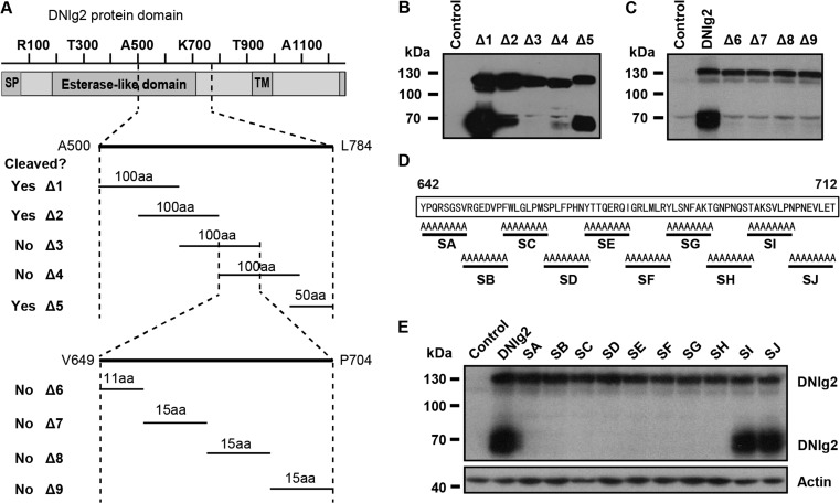
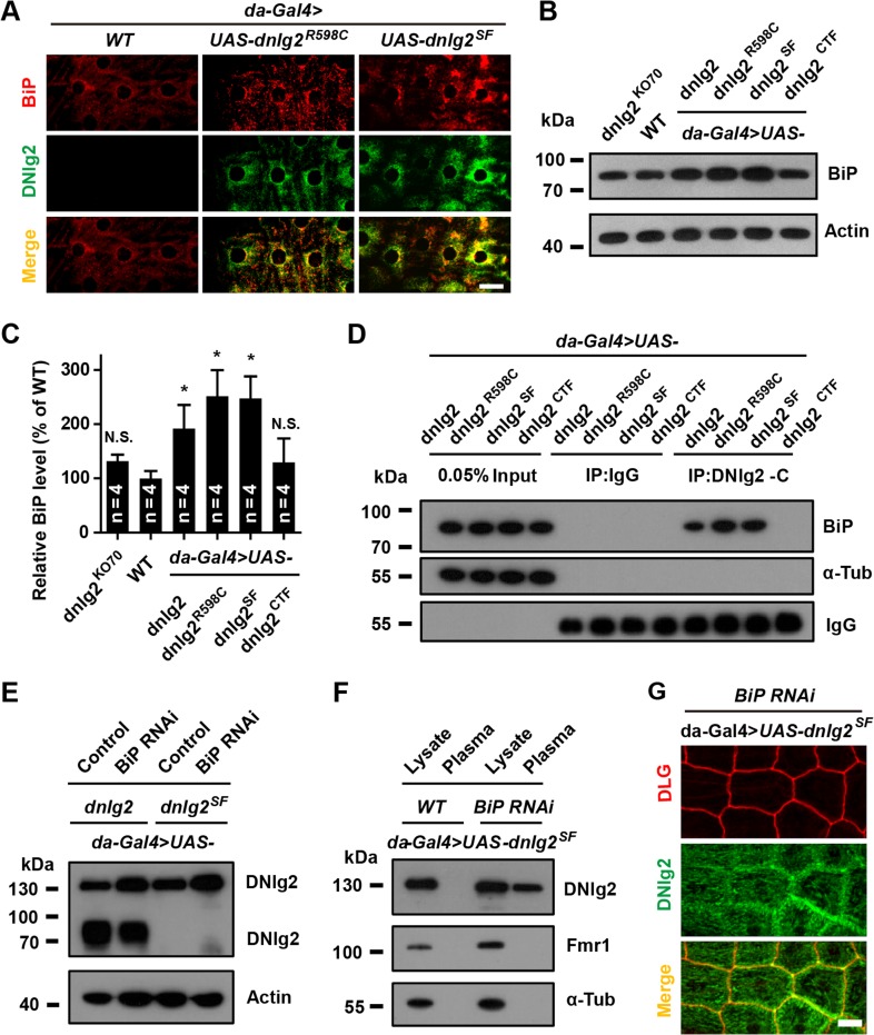
Neuroligins (Nlgs) are transmembrane cell adhesion molecules playing essential roles in synapse development and function. Genetic mutations in neuroligin genes have been linked with some neurodevelopmental disorders such as autism. These mutated Nlgs are mostly retained in the endoplasmic reticulum (ER). However, the mechanisms underlying normal Nlg maturation and trafficking have remained largely unknown. Here, we found that Drosophila neuroligin 2 (DNlg2) undergoes proteolytic cleavage in the ER in a variety of Drosophila tissues throughout developmental stages. A region encompassing Y642-T698 is required for this process. The immature non-cleavable DNlg2 is retained in the ER and non-functional. The C-terminal fragment of DNlg2 instead of the full-length or non-cleavable DNlg2 is able to rescue neuromuscular junction defects and GluRIIB reduction induced by dnlg2 deletion. Intriguingly, the autism-associated R598C mutation in DNlg2 leads to similar marked defects in DNlg2 proteolytic process and ER export, revealing a potential role of the improper Nlg cleavage in autism pathogenesis. Collectively, our findings uncover a specific mechanism that controls DNlg2 maturation and trafficking via proteolytic cleavage in the ER, suggesting that the perturbed proteolytic cleavage of Nlgs likely contributes to autism disorder.

Keywords: autism; maturation; neuroligin; proteolytic cleavage; trafficking.


Figures

Figure 1
Figure 1
Proteolytic cleavage of DNlg2. (A) Immunoblotting analysis of Gal4/Tub-Gal80ts-controlled DNlg2-overexpressing muscle cells. Entrainment temperature and incubated time (h) are shown at the top. (B) Immunoblotting analysis of WT Drosophila samples at different developmental stages (1−4: embryonic 0−6 h, 6−12 h, 12−18 h, 18−24 h; 5−8: larval of 1st, 2nd, early 3rd, late 3rd instar; 9−11: pupal head after 24, 48, 72 h pupation; 12−14: adult head 1 day, 3 days, 5 days). (C) Immunoblotting analysis of dnlg2 mutant, WT, elav-Gal4 > UAS-dnlg2, da-Gal4 > UAS-dnlg2, gmr-Gal4 > UAS-dnlg2, and repo-Gal4 > UAS-dnlg2 fly heads. (D) Immunoblotting analysis of S2 cells transfected with pAc5.1-dnlg2 plasmid after 24, 36, 48, or 72 h. (E) Immunoblotting analysis of protease inhibitor (PI)-treated samples. DNlg2 proteins were overexpressed with the Gal4/Tub-Gal80ts expression system for 37 h. Then dissected muscle cells were incubated at 25°C with or without PI cocktail for 6 or 12 h. (F) Immunoblotting analysis of human 293T cells transfected with 0, 0.5, 1, or 2 μg pcDNA3.1-dnlg2 plasmid. The rabbit anti-DNlg2CTF antibodies were used.
Figure 2
Figure 2
Proteolytic cleavage of DNlg2 occurs in the ER. (A) Immunoblotting analysis of samples collected from purified plasma membrane fractions. Blots were probed with antibodies against DNlg2, Fmr1, and α-Tubulin. Lysate means whole-cell fractions. Plasma means purified plasma membrane fractions. OE means DNlg2-overexpressing cells. (B) Immunoblotting analysis of samples collected from a 10%–30% density gradient separation prepared from adult WT heads. Blots were probed with antibodies against DNlg2 intracellular domain, the Golgi protein Syn16, and the ER protein BiP. (C) DNlg2 proteins were overexpressed and immunoprecipitated, and then digested with or without 2.5 μg/ml trypsin for 1, 3, 6, or 10 min. Samples were then followed by immunoblotting with anti-DNlg2 antibody. The rabbit anti-DNlg2CTF antibodies were used.
Figure 3
Figure 3
Y642−T698 region is required for DNlg2 cleavage. (A) Schematic diagram of DNlg2 structure. Positions of representative amino acids are shown above. Amino acid lengths and positions of deleted DNlg2 fragments are shown below (Δ1: A500−D599, Δ2: F550−V649, Δ3: A600−A699, Δ4: A650−E749, Δ5: N734−L784, Δ6: V649−G659, Δ7: L660−E674, Δ8: R675−A689, and Δ9: K690−P704). (B) Immunoblotting analysis of S2 cells transfected with large deleted DNlg2 fractions. (C) Immunoblotting analysis of S2 cells transfected with small deleted DNlg2 sequences. (D) Schematic depiction of protein sequences of alanine substitution mutant DNlg2. (E) Immunoblotting analysis of S2 cells transfected with alanine substitution mutant DNlg2 constructs. The rabbit anti-DNlg2CTF antibodies were used in B, C, and E.
Figure 4
Figure 4
R598C mutant DNlg2 shows abnormal proteolytic cleavage and trafficking. (A) Multiple sequence alignment of Homo sapiens (Hs), Mus musculus (Ms), and Drosophila melanogaster (Dm) Nlgs. Red star points at the Hs NLG3 R541 amino acid. Hs NLG1 (NP_055747.1), Hs NLG 2 (NP_065846), Hs NLG 3 (NP_061850), Hs NLG 4 (NP_065793.1), Mm Nlg1 (NP_619607.2), Mm Nlg2 (NP_942562.2), Mm Nlg3 (NP_766520.2), Mm Nlg4 (ABS19580), Dm Nlg1 (NP_731172), Dm Nlg2 (NP_523496.1), Dm Nlg3 (NP_001036685.2),  and Dm Nlg4 (NP_001163661.1). (B) Confocal images of larval brains stained with mouse anti-DNlg26D5 (green) and rabbit anti-DVGLUT (red). Genotypes were WT, elav-Gal4 > UAS-dnlg2, and elav-Gal4 > UAS-dnlg2R598C. Scale bar, 50 μm. Arrows point at synapse area and arrowheads point at soma site. (C) Quantification of relative DNlg2 protein amount on synapse of each genotype. (D) Quantification of relative DNlg2 protein amount in soma of each genotype. (E) Immunoblotting analysis of DNlg2 in third instar larval muscle cells with shorter (30 sec) and longer (3 min) exposure time. Genotypes were da-Gal4 > WT, da-Gal4 > UAS-dnlg2, and da-Gal4 > UAS-dnlg2R598C. The rabbit anti-DNlg2CTF antibodies were used. (F) Quantification of the 70 kDa DNlg2 proportion in total proteins. Error bar indicates SEM. **P < 0.01, *P < 0.05.
Figure 5
Figure 5
CTF but not non-cleavable mutant DNlg2 is transported to dendrites or cell surfaces. (A) Schematic representation of mutant or N-terminal deleted DNlg2. (B) Immunoblotting analysis of dnlg2 mutant, WT, and UAS-dnlg2, UAS-dnlg2R598C, UAS-dnlg2SF, UAS-dnlg2CTF flies with da-Gal4. Third instar larval muscle cells were used. The rabbit anti-DNlg2CTF antibodies were used. (C) Confocal images showing DNlg2 distributions in pdf-Gal4 > WT, pdf-Gal4 > UAS-dnlg2, pdf-Gal4 > UAS-dnlg2R598C, pdf-Gal4 > UAS-dnlg2SF, and pdf-Gal4 > UAS-dnlg2CTF adult brain dendrite region, labeled with mouse anti-DNlg26D5 (red) and rabbit anti-GFP (green). Scale bar, 10 μm. (D) Confocal images showing DNlg2 distributions of da-Gal4 > WT, da-Gal4 > UAS-dnlg2, da-Gal4 > UAS-dnlg2R598C, da-Gal4 > UAS-dnlg2SF, and da-Gal4 > UAS-dnlg2CTF epithelial cells, labeled with rabbit anti-DNlg2CTF (green) and mouse anti-DLG (red). Scale bar, 25 μm.
Figure 6
Figure 6
Full-length DNlg2 is retained in the ER through binding to BiP. (A) Confocal images showing non-cleavable DNlg2 (da-Gal4 > UAS-dnlg2 and da-Gal4 > UAS-dnlg2R598C) co-localized with BiP. Dissected epithelial cells were co-stained with rabbit anti-DNlg2CTF (green) and rat anti-BiP (red, showing the ER). Scale bar, 25 μm. (B) Immunoblotting analysis of dnlg2 mutant, WT, and da-Gal4 > UAS-dnlg2, da-Gal4 > UAS-dnlg2R598C, da-Gal4 > UAS-dnlg2SF, da-Gal4 > UAS-dnlg2CTF flies with anti-BiP. (C) Quantification of relative BiP expression level in each genotype. (D) Co-immunoprecipitation of DNlg2 and BiP in vivo. (E) Immunoblotting analysis of da-Gal4 overexpressing UAS-dnlg2 and UAS-dnlg2SF at WT or BiP RNAi background with rabbit anti-DNlg2CTF. (F) Immunoblotting analysis of da-Gal4 > UAS-dnlg2SF and da-Gal4 > UAS-BiP-RNAi,UAS-dnlg2SF with rabbit anti-DNlg2CTF. Samples were purified with plasma−membrane extraction kit. (G) Confocal images of UAS-dnlg2SF-overexpressing flies under BiP knockdown background with da-Gal4. Dissected epithelial cells were co-stained with rabbit anti-DNlg2CTF (green) and mouse anti-DLG antibody (red). Scale bar, 20 μm.
Figure 7
Figure 7
The 70 kDa DNlg2 promotes NMJ development. (A) Confocal images of NMJ6/7 from abdominal segment 2 of third instar larvae labeled with rabbit anti-HRP (green) and mouse anti-DLG (red), showing bouton phenotypes in WT, dnlg2KO70, and rescue lines with UAS-dnlg2, UAS-dnlg2R598C, UAS-dnlg2SF, UAS-dnlg2CTF derived by da-Gal4. Scale bar, 25 μm. (B) Quantification of total bouton number at NMJ6/7, adjusted to WT bouton number. (C) Confocal images of synaptic boutons at NMJ4 labeled with rabbit anti-GluRIIB (green) and mouse anti-BRP (red), showing GluRIIB signal phenotypes in WT, dnlg2KO70, and rescue lines with UAS-dnlg2, UAS-dnlg2R598C, UAS-dnlg2SF, UAS-dnlg2CTF derived by da-Gal4. Scale bar, 5 μm. (D) Quantification of GluRIIB signal intensity with 3D reconstructed confocal images.

Similar articles

Cited by

References

    1. Banovic D., Khorramshahi O., Owald D., et al. (2010). Drosophila neuroligin 1 promotes growth and postsynaptic differentiation at glutamatergic neuromuscular junctions. Neuron 66, 724–738. - PubMed
    1. Bischof J., Maeda R.K., Hediger M., et al. (2007). An optimized transgenesis system for Drosophila using germ-line-specific phiC31 integrases. Proc. Natl Acad. Sci. USA 104, 3312–3317. - PMC - PubMed
    1. Chubykin A.A., Atasoy D., Etherton M.R., et al. (2007). Activity-dependent validation of excitatory versus inhibitory synapses by neuroligin-1 versus neuroligin-2. Neuron 54, 919–931. - PMC - PubMed
    1. Comoletti D., De Jaco A., Jennings L.L., et al. (2004). The Arg451Cys-neuroligin-3 mutation associated with autism reveals a defect in protein processing. J. Neurosci. 24, 4889–4893. - PMC - PubMed
    1. Conant K., Allen M., and Lim S.T. (2015). Activity dependent CAM cleavage and neurotransmission. Front. Cell. Neurosci. 9, 305. - PMC - PubMed

Publication types

MeSH terms