. 2017 Jul 1;216(1):82-91.
doi: 10.1093/infdis/jix223.

Pathological Role of Anti-CD4 Antibodies in HIV-Infected Immunologic Nonresponders Receiving Virus-Suppressive Antiretroviral Therapy

Affiliations

Pathological Role of Anti-CD4 Antibodies in HIV-Infected Immunologic Nonresponders Receiving Virus-Suppressive Antiretroviral Therapy

Zhenwu Luo et al. J Infect Dis. .

Abstract

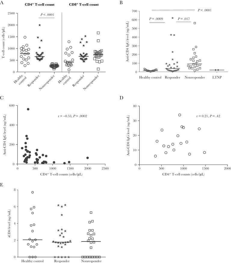
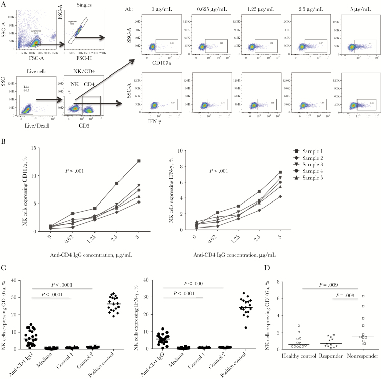
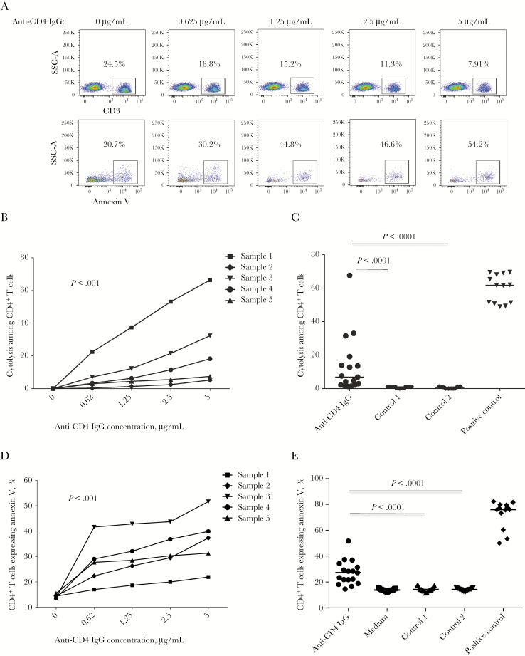
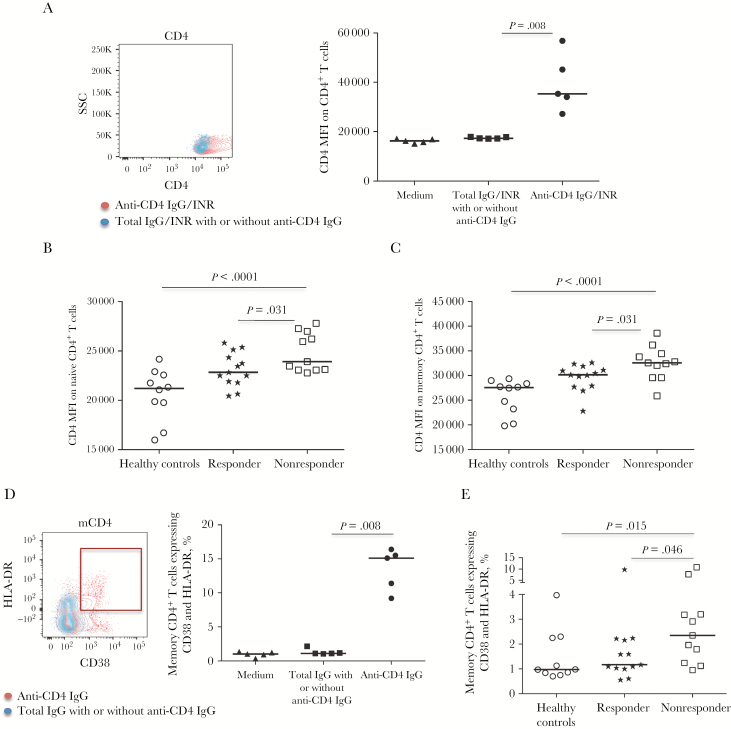
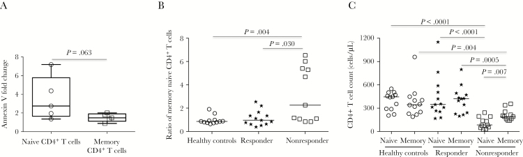
Increased mortality and morbidity occur among human immunodeficiency virus (HIV)-infected patients in whom CD4+ T-cell counts do not increase despite viral suppression with antiretroviral therapy (ART). Here we identified an underlying mechanism. Significantly elevated plasma levels of anti-CD4 immunoglobulin G (IgG) were found in HIV-positive immunologic nonresponders (ie, HIV-positive individuals with CD4+ T-cell counts of ≤350 cells/μL), compared with levels in HIV-positive immunologic responders (ie, HIV-positive individuals with CD4+ T-cell counts of ≥500 cells/μL) and healthy controls. Higher plasma level of anti-CD4 IgG correlated with blunted CD4+ T-cell recovery. Furthermore, purified anti-CD4 IgG from HIV-positive immunologic nonresponders induced natural killer (NK) cell-dependent CD4+ T-cell cytolysis and apoptosis through antibody-dependent cell-mediated cytotoxicity (ADCC) in vitro. We also found that anti-CD4 IgG-mediated ADCC exerts greater apoptosis of naive CD4+ T cells relative to memory CD4+ T cells. Consistently, increased frequencies of CD107a+ NK cells and profound decreases of naive CD4+ T cells were observed in immunologic nonresponders as compared to responders and healthy controls ex vivo. These data indicate that autoreactive anti-CD4 IgG may play an important role in blunted CD4+ T-cell reconstitution despite effective ART.

Keywords: B cells; HIV; antibody responses; autoreactive anti-CD4 antibodies.


Figures

Figure 1.
Figure 1.
Increased plasma anti-CD4 immunoglobulin G (IgG) levels in aviremic antiretroviral therapy (ART)–recipient immunologic nonresponders. A, Median absolute numbers of peripheral CD4+ and CD8+ T cells, assessed by flow cytometry, in healthy controls, responders, and nonresponders. B, Median plasma levels of anti-CD4 IgG in healthy controls, responders, nonresponders, and long-term nonprogressors (LTNPs). C and D, Correlations between plasma levels of anti-CD4 IgG and peripheral CD4+ T-cell counts in all ART-recipient aviremic human immunodeficiency virus (HIV)–infected subjects (C) and healthy controls (D). E, Median plasma levels of soluble CD4 (sCD4) antigen in healthy controls, responders, and nonresponders. Statistical analyses were performed using the Mann-Whitney U test (unpaired).
Figure 2.
Figure 2.
Anti-CD4 antibody-dependent natural killer (NK) cell activation. CD4+ T cells were cultured with purified anti-CD4 immunoglobulin G (IgG) from nonresponders or control antibodies and cocultured with NK cells at a ratio of 1:1. Intracellular CD107a and interferon γ (IFN-γ) expression in NK cells was analyzed by flow cytometry. A, Dot plots from a representative donor show CD107a and IFN-γ expression in NK cells in response to different concentrations of anti-CD4 antibodies from nonresponders. B, Percentages of NK cells expressing IFN-γ and CD107a in response to different concentrations of anti-CD4 IgG from 5 different nonresponders in vitro. Analyses were performed by the Friedman paired nonparametric test. C, Median percentages of NK cells expressing IFN-γ and CD107a in a mixed culture of CD4+ T cells and NK cells in the presence of anti-CD4 IgG from nonresponders, anti-CD4 IgG from nonresponders pretreated with sCD4 (control 1), anti-CD4 IgG–depleted total IgG from nonresponders (control 2), and zanolimumab (positive control) at 5 μg/mL in vitro. Analyses were performed using the Mann-Whitney U test (unpaired). D, Median percentages of NK cells expressing CD107a in healthy controls, responders, and nonresponders ex vivo. Analyses were performed using the Mann-Whitney U test (unpaired). FSC, forward scatter; SSC, side scatter.
Figure 3.
Figure 3.
Anti-CD4 antibody dependent natural killer (NK) cell–mediated cytolysis to primary CD4+ T cells. CD4+ T cells were cultured with purified anti-CD4 immunoglobulin G (IgG) and control antibodies and cocultured with NK cells at a ratio of 1:3 in vitro, and the CD4+ T-cell cytolysis percentage was analyzed by flow cytometry. A, Dot plots from 1 representative donor were showing CD4+ T-cell frequencies and annexin V binding in response to different concentrations of anti-CD4 IgG from nonresponders. B, Percentages of CD4+ T cells undergoing cytolysis in response to different concentrations of anti-CD4 IgG from 5 different nonresponders through antibody-dependent cell-mediated cytotoxicity (ADCC). Analyses were performed by the Friedman paired nonparametric test. C, Median percentages of CD4+ T cells undergoing cytolysis in response to anti-CD4 IgG from nonresponders and controls at 5 μg/mL in vitro. Analyses were performed using the Mann-Whitney U test (unpaired). D, Percentages of CD4+ T cells undergoing apoptosis in response to different concentrations of anti-CD4 IgG from 5 different nonresponders through ADCC. Analyses were performed by the Friedman paired nonparametric test. E, Percentages of CD4+ T cells undergoing apoptosis in response to anti-CD4 IgG from nonresponders and controls at 5 μg/mL in vitro. Analyses were performed using the Mann-Whitney U test (unpaired). SSC, side scatter.
Figure 4.
Figure 4.
CD4+ T-cell activation in response to purified anti-CD4 immunoglobulin G (IgG) from immunologic nonresponders. A, Overlapped dot plots (1 representative donor) and the median CD4 mean fluorescence intensity (MFI) expression on CD4+ T cells treated with anti-CD4 IgG (red) and anti-CD4 IgG–depleted total IgG (blue) in vitro. B and C, Median CD4 MFI expression on naive CD4+ T cells (B) and memory CD4+ T cells (C) in healthy controls, responders, and nonresponders ex vivo. D, Overlapped dot plots (1 representative donor) and median percentages of CD38 and HLA-DR coexpression on memory CD4+ T cells treated with anti-CD4 IgG (red) and anti-CD4 IgG–depleted total IgG in vitro. E, Median percentages of CD38 and HLA-DR coexpression on memory CD4+ T cells in healthy controls, responders, and nonresponders ex vivo. Analyses were performed using the Mann-Whitney U test (unpaired). SSC, side scatter.
Figure 5.
Figure 5.
Memory and naive CD4+ T cells. A, Comparison of apoptotic induction between naive CD4+ and memory CD4+ T cells by purified anti-CD4 immunoglobulin G (IgG) from plasma of 5 different nonresponders in antibody-dependent cell-mediated cytotoxicity assays in vitro. Data are median fold change (interquartile range) from anti-CD4 IgG to anti-CD4 IgG–depleted total IgG. B, Median ratio of memory CD4+ T cells to naive CD4+ T cells in healthy controls, responders, and nonresponders ex vivo. C, Median absolute counts of memory CD4+ and naive CD4+ T cells in healthy controls, responders, and nonresponders ex vivo. Analyses were performed using the Mann-Whitney U test (unpaired).

References

    1. Mocroft A, Vella S, Benfield TL, et al. Changing patterns of mortality across Europe in patients infected with HIV-1. EuroSIDA Study Group. Lancet 1998; 352:1725–30. - PubMed
    1. Battegay M, Nüesch R, Hirschel B, Kaufmann GR. Immunological recovery and antiretroviral therapy in HIV-1 infection. Lancet Infect Dis 2006; 6:280–7. - PubMed
    1. May MT, Sterne JA, Costagliola D, et al. ; Antiretroviral Therapy (ART) Cohort Collaboration. HIV treatment response and prognosis in Europe and North America in the first decade of highly active antiretroviral therapy: a collaborative analysis. Lancet 2006; 368:451–8. - PubMed
    1. Kelley CF, Kitchen CM, Hunt PW, et al. Incomplete peripheral CD4+ cell count restoration in HIV-infected patients receiving long-term antiretroviral treatment. Clin Infect Dis 2009; 48:787–94. - PMC - PubMed
    1. Lederman MM, Calabrese L, Funderburg NT, et al. Immunologic failure despite suppressive antiretroviral therapy is related to activation and turnover of memory CD4 cells. J Infect Dis 2011; 204:1217–26. - PMC - PubMed

Publication types

MeSH terms