. 2017 Jun 7;139(22):7494-7503.
doi: 10.1021/jacs.7b01158. Epub 2017 May 23.

Identification of Intrahelical Bifurcated H-Bonds as a New Type of Gate in K+ Channels

Affiliations

Identification of Intrahelical Bifurcated H-Bonds as a New Type of Gate in K+ Channels

Oliver Rauh et al. J Am Chem Soc. .

Abstract

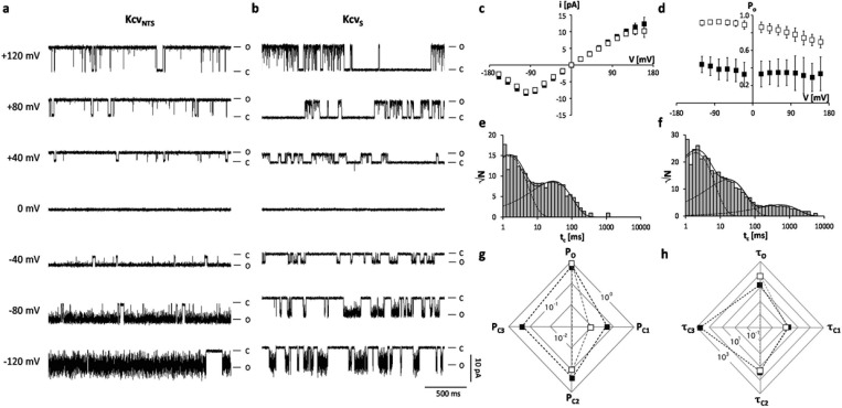
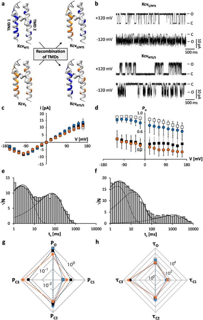
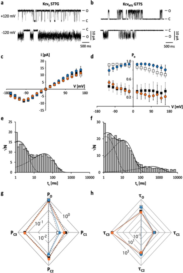
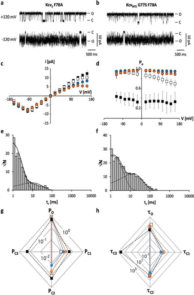
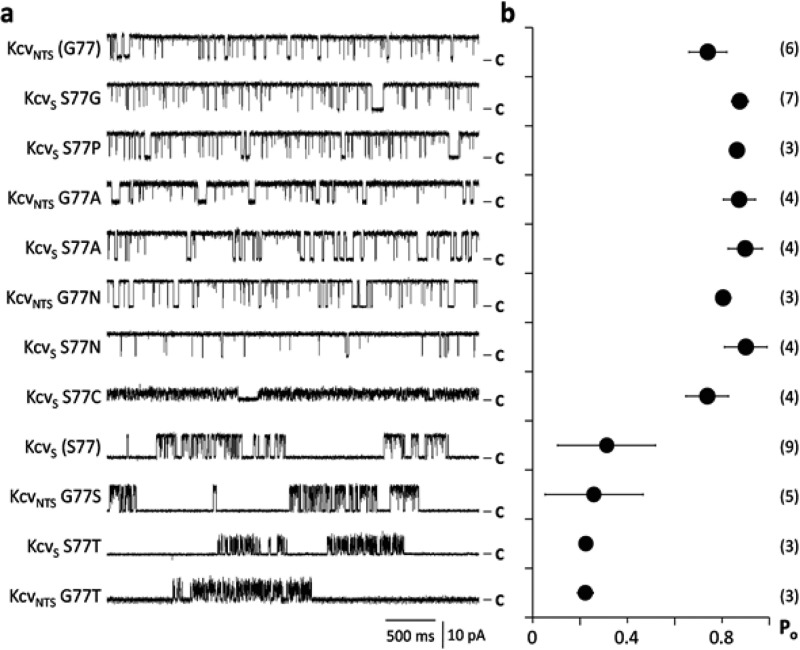
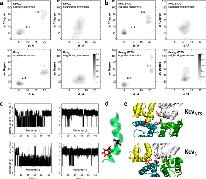
Gating of ion channels is based on structural transitions between open and closed states. To uncover the chemical basis of individual gates, we performed a comparative experimental and computational analysis between two K+ channels, KcvS and KcvNTS. These small viral encoded K+ channel proteins, with a monomer size of only 82 amino acids, resemble the pore module of all complex K+ channels in terms of structure and function. Even though both proteins share about 90% amino acid sequence identity, they exhibit different open probabilities with ca. 90% in KcvNTS and 40% in KcvS. Single channel analysis, mutational studies and molecular dynamics simulations show that the difference in open probability is caused by one long closed state in KcvS. This state is structurally created in the tetrameric channel by a transient, Ser mediated, intrahelical hydrogen bond. The resulting kink in the inner transmembrane domain swings the aromatic rings from downstream Phes in the cavity of the channel, which blocks ion flux. The frequent occurrence of Ser or Thr based helical kinks in membrane proteins suggests that a similar mechanism could also occur in the gating of other ion channels.


Conflict of interest statement

The authors declare no competing financial interest.

Figures

Figure 1
Figure 1
Chlorovirus encoded K+ channels. (a) Cartoon representations of a snapshot of KcvS taken from MD simulation at 80 ns of the production run. Left: Side view of the channel (two opposing monomer units). Right: view from bottom to top of the full tetrameric channel. Potassium ions are shown as red spheres, and F78 as explicit side chains in red. (b) Sequence alignment of three viral encoded K+ channels. The position of the transmembrane domains TMD1 and TMD2 as well as the pore helix are indicated by bars. The sequences are 86% identical; amino acid differences are highlighted in gray.
Figure 2
Figure 2
The difference in open probability of two similar K+ channels originates from a long closed time, which is absent in KcvNTS and present in KcvS. (a,b) Characteristic single channel fluctuations of KcvNTS and KcvS at different voltages in a planar lipid bilayer. The closed (C) and open (O) levels are indicated along the current traces. (c) Mean single channel i/V relations (±sd) of KcvNTS (open squares) and KcvS (filled squares) from 6 and 9 independent recordings, respectively. (d) Mean open probabilities (±sd) of the two channels from 6 and 9 independent recordings. Exemplary closed dwell times at +120 mV for KcvNTS (e) and KcvS (f). The data in e can be fitted with two exponentials while the data in f require a third exponential. The long-lived closed state in f is absent in e. The multiple kinetic variables of the two channels including the probabilities (P) for occupying the open state (O) and the closed states (C1–C3) (g) as well as mean lifetimes (in ms) of the open state (τo) and of the three closed states (τc1–τc3) (h) are presented in two-dimensional radar plots. The probabilities of occupancy and mean lifetimes were calculated from three independent 5 min recordings. The symbols in d, g and h correspond to those in c.
Figure 3
Figure 3
The KcvS long closed time is related to TMD2. (a) Chimeras were constructed by swapping transmembrane domains between KcvNTS (in blue) and KcvS (orange), for illustration purposes only. (b) Characteristic single channel fluctuations of chimeras KcvS/NTS and KcvNTS/S at ±120 mV in planar lipid bilayers. (c) Mean single channel i/V relations (±sd) of KcvNTS (open squares), KcvS (filled squares), KcvS/NTS (blue circles, n = 9) and KcvNTS/S (orange circles, n = 3). (d) Mean open probabilities (±sd) of wt channels and chimeras (n = 9 and n = 3 for KcvS/NTS and KcvNTS/S, respectively). Exemplary closed dwell times at +120 mV for KcvS/NTS (e) and KcvNTS/S (f). The data in e can be fitted with two exponentials while the data in f require a third exponential. Radar plots for probabilities (P) of the wt channels and the chimeras for occupying the open state (O) and the closed states (C1–C3) (g) and of mean lifetimes (in ms) of the open state (τo) and of the three closed states (τc1–τc3) (h). The probabilities of occupancy and mean lifetimes were calculated from three independent 5 min recordings. The symbols in d, g and h correspond to those in c.
Figure 4
Figure 4
The difference of KcvNTS and KcvS in position 77 is responsible for the absence or presence of a long closed time. Characteristic single channel fluctuations of mutant KcvS S77G (a) and KcvNTS G77S (b) at ±120 mV in planar lipid bilayers. (c) Mean single channel i/V relations (±sd) of KcvNTS (open squares), KcvS (filled squares), KcvNTS G77S (orange circles, n = 5) and KcvS S77G (blue circles, n = 3). (d) Mean open probabilities (±sd) of wt channels and mutants (n = 5 and n = 3 for KcvNTS G77S and KcvS S77G, respectively). Exemplary closed dwell times at +120 mV for KcvS S77G (e) and KcvNTS G77S (f). The data in e can be fitted with two exponentials while the data in f again require a third exponential. Radar plots for probabilities (P) of the wt channels and mutants for occupying the open state (O) and the closed states (C1–C3) (g) and for mean lifetimes (in ms) of the open state (τo) and of the three closed states (τc1–τc3) (h). The probabilities of occupancy and mean lifetimes were calculated from three independent 5 min recordings. The symbols in d, g and h correspond to those in c.
Figure 5
Figure 5
The aromatic AA F78 functions in combination with the upstream AA as a gate for the long closed time. Characteristic single channel fluctuations of mutant KcvS F78A (a) and KcvNTS G77S F78A (b) at ±120 mV in planar lipid bilayers. (c) Mean single channel i/V relations (±sd) of KcvNTS (open squares), KcvS (filled squares), KcvS F78A (orange circles, n = 4) and KcvNTS G77S F78A (blue circles, n = 7). (d) Mean open probabilities (±sd) of wt channels and mutants (n = 4 and n = 7 for KcvS F78A and KcvNTS G77S F78A, respectively). Exemplary closed dwell times at +120 mV for KcvS F78A (e) and KcvNTS G77S F78A (f). The data in e and f can now be fitted with two exponentials. Radar plots for probabilities (P) of the wt channels and mutants for occupying the open state (O) and the closed states (C1–C3) (g) and for mean lifetimes (in ms) of the open state (τo) and of the three closed states (τc1–τc3) (h). The mean lifetimes were calculated from three independent 5 min recordings. The symbols in d, g and h correspond to those in c.
Figure 6
Figure 6
The AA S77 is responsible for the gate with the long closed time. (a) Characteristic single channel fluctuations at +120 mV of KcvS and KcvNTS and mutants in which the position 77 in both channels was exchanged with an AA with a different flavor. (b) Mean open probabilities (±sd) of wt channels and mutants. The number of independent measurements is shown in brackets. Data in b correspond to the constructs on the left in a. Only AAs Ser and Thr in position 77 generate a low open probability with a long lasting closed time.
Figure 7
Figure 7
Dynamics of TMD2 in KcvS/KcvNTS and mutants. (a) Distribution of distance-angle pairs characterizing π-stack geometries in KcvNTS (top row) and KcvS (bottom row) averaged over all F78 pairs in opposite (left column) and neighboring (right column) monomers. The geometric criteria are shown Figure S5. Numbers in the left panel denote different monomers. (b) Analogous data for KcvS S77G and KcvNTS G77S mutants, demonstrating the transfer of π-stack characteristics upon mutating the key residues. (c) Time series of the distances between the carbonyl oxygen of I73 and the hydroxyl hydrogen of S77 for all four monomers. (d) Schematic representation of a hydrogen bond formed between the amide oxygen of I73 and the hydroxyl hydrogen of S77 within monomer 3 of KcvS supplemented by orientation of F78 (snapshot taken at 80 ns). (e) Snapshots of F78 (red) in tetramer of KcvNTS (top) and KcvS (bottom) representing a wider and a narrow translocation pathway corresponding to statistics shown in a.
Figure 8
Figure 8
The two channels KcvS and KcvNTS have a different orientation of the C-terminus. Characteristic single channel fluctuations at +120 mV of KcvS W82C (a) and KcvNTS W82C (b) in absence (−DTT) and presence of 5 mM DTT (+DTT) in bath solution. The prevailing open (O) and closed (C) levels are indicated along the traces.

Similar articles

Cited by

References

    1. Hille B.Ion Channels of Excitable Membranes, 3rd ed.; Sinauer Associates Inc.: Sunderland, 2001.
    1. Cordero-Morales J. F.; Cuello L. G.; Zhao Y.; Jogini V.; Cortes D.; Roux B.; Perozo E. Nat. Struct. Mol. Biol. 2006, 13, 311.10.1038/nsmb1069. - DOI - PubMed
    1. Thompson A. N.; Posson D. J.; Rarsa P. V.; Nimigean C. M. Proc. Natl. Acad. Sci. U. S. A. 2008, 105, 6900.10.1073/pnas.0800873105. - DOI - PMC - PubMed
    1. Tayefeh S.; Kloss T.; Kreim M.; Gebhardt M.; Baumeister D.; Hertel B.; Richter C.; Schwalbe H.; Moroni A.; Thiel G.; Kast S. M. Biophys. J. 2009, 96, 485.10.1016/j.bpj.2008.09.050. - DOI - PMC - PubMed
    1. Thiel G.; Baumeister D.; Schroeder I.; Kast S. M.; Van Etten J. L.; Moroni A. Biochim. Biophys. Acta, Biomembr. 2011, 1808, 580.10.1016/j.bbamem.2010.04.008. - DOI - PubMed

Publication types

Substances

LinkOut - more resources