. 2017 May 12;12(1):38.
doi: 10.1186/s13024-017-0179-7.

Anterograde monosynaptic transneuronal tracers derived from herpes simplex virus 1 strain H129

Affiliations

Anterograde monosynaptic transneuronal tracers derived from herpes simplex virus 1 strain H129

Wen-Bo Zeng et al. Mol Neurodegener. .

Abstract

Background: Herpes simplex virus type 1 strain 129 (H129) has represented a promising anterograde neuronal circuit tracing tool, which complements the existing retrograde tracers. However, the current H129 derived tracers are multisynaptic, neither bright enough to label the details of neurons nor capable of determining direct projection targets as monosynaptic tracer.

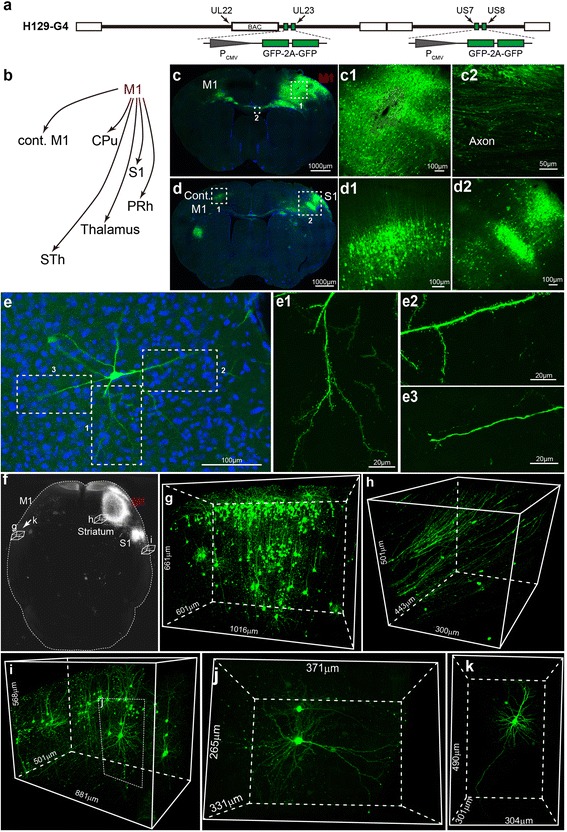
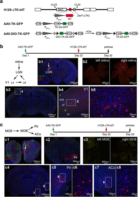
Methods: Based on the bacterial artificial chromosome of H129, we have generated a serial of recombinant viruses for neuronal circuit tracing. Among them, H129-G4 was obtained by inserting binary tandemly connected GFP cassettes into the H129 genome, and H129-ΔTK-tdT was obtained by deleting the thymidine kinase (TK) gene and adding tdTomato coding gene to the H129 genome. Then the obtained viral tracers were tested in vitro and in vivo for the tracing capacity.

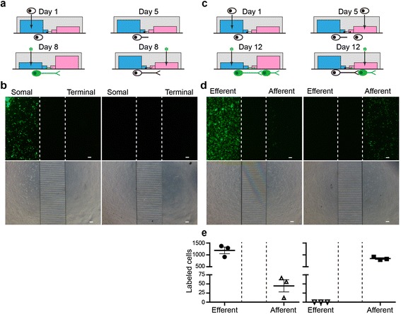
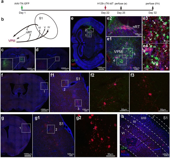
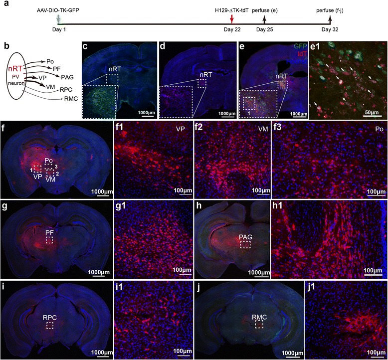
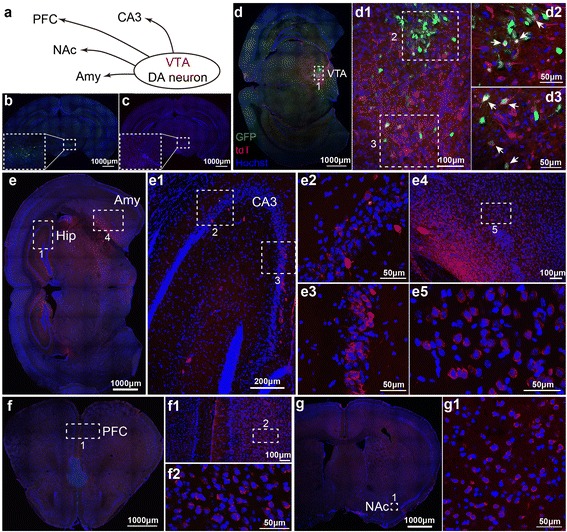
Results: H129-G4 is capable of transmitting through multiple synapses, labeling the neurons by green florescent protein, and visualizing the morphological details of the labeled neurons. H129-ΔTK-tdT neither replicates nor spreads in neurons alone, but transmits to and labels the postsynaptic neurons with tdTomato in the presence of complementary expressed TK from a helper virus. H129-ΔTK-tdT is also capable to map the direct projectome of the specific neuron type in the given brain regions in Cre transgenic mice. In the tested brain regions where circuits are well known, the H129-ΔTK-tdT tracing patterns are consistent with the previous results.

Conclusions: With the assistance of the helper virus complimentarily expressing TK, H129-ΔTK-tdT replicates in the initially infected neuron, transmits anterogradely through one synapse, and labeled the postsynaptic neurons with tdTomato. The H129-ΔTK-tdT anterograde monosynaptic tracing system offers a useful tool for mapping the direct output in neuronal circuitry. H129-G4 is an anterograde multisynaptic tracer with a labeling signal strong enough to display the details of neuron morphology.

Keywords: Anterograde; H129; H129 strain; H129-G4; Herpes simplex virus type 1 (HSV-1); Monosynaptic; Multisynaptic; Neuronal tracer; tdT; ΔTK.


Figures

Fig. 1
Fig. 1
H129 derived tracer H129-G4 (a) The schematic diagram of H129-G4 genome. Two binary GFP elements were inserted into the genome of H129 bacterial artificial chromosome (H129-BAC) at the indicated position. b Schema of the simplified M1 projection pathways. The M1 pathway has been simplified and only the representative M1 projection targets are displayed. M1, primary motor cortex; cont. M1, contralateral M1; S1, primary somatosensory cortex; PRh, perirhinal cortex; STh, subthalamic nucleus; CPu, caudate putamen. cd Representative tracing result of H129-G4. H129-G4 was injected into M1 of wild-type C57BL/6 mice, and representative images of coronal brain sections were obtained at 4 days post infection (dpi). Representative regions are shown, and the boxed areas are presented in the right panels with a higher magnification. e A representative H129-G4 labeled single neuron. A representative GFP-labeled neuron in PRh is shown, and the magnified images of the dendritic segments with individual spines (e1 and e2) and the axon (e3) are presented in the right panels. fk Combination of fMOST and H129-G4 tracing. The mouse brain obtained at 4 dpi was further processed to fMOST imaging. The 3D image of the whole brain was reconstructed (f). Representative brain regions, including the contralateral S1 (g), striatum (h) and the ipsilateral S1 (i), are shown in details. Representative single neuron images at the ipsilateral S1 (j) and the contralateral S1 (k) are also presented
Fig. 2
Fig. 2
Invasion and transmission of H129-G4 in vitro (ab) Invasion of H129-G4. Freshly isolated fetal mouse hippocampal and cortical neurons were seeded into one chamber of the microfluidic plate, and the first 24 h culture was termed as Day 1. H129-G4 was added at Day 8 into either the soma (blue) or axon terminal chamber (red) to a final concentration of 1 × 107 pfu/ml (a). Images of GFP signal (upper panel) and phase contrast (lower panel) were obtained at 24 h post infection (hpi). The represent results from 3 microchannel plates are shown (b). The dotted lines indicate the borders between chambers and the microchannels. Scale bar = 100 μm (ce) Transmission direction of H129-G4. Neurons were sequentially plated into the two chambers at Day 1 and Day 5, then H129-G4 was added at Day 12 into either the efferent (blue) or afferent chamber (red) to a final concentration of 1 × 107pfu/ml (c). Images of GFP signal (upper panel) and phase contrast (lower panel) were obtained at 24hpi. The representative results from 3 microfluidic plates are shown (d), and the quantitative analysis were performed by counting the GFP labeled cell mounts in different chambers (e). The dotted lines indicate the borders between chambers and the microchannel. Scale bar = 100 μm
Fig. 3
Fig. 3
In vivo transmission of H129-ΔTK-tdT (a) Schematic genome diagrams of H129-ΔTK-tdT and the helper viruses. H129-ΔTK-tdT was generated by replacing the thymidine kinase gene (TK, UL23) of H129-BAC with CMV promoter controlled tdTomato (tdT) gene and the selection marker of zeocin resistance gene (ZeoR). The helper viruses of AAV-TK-GFP and AAV-DIO-TK-GFP express TK and GFP under the control of EF1α promoter either in the Cre-independent or -dependent manner, respectively. bc Transmission of H129-ΔTK-tdT tracing system in vivo. AAV-TK-GFP and H129-ΔTK-tdT were sequentially injected into LGN (b) and MOB (c) with the dose following the table described in Materials and methods. Representative upstream or downstream brain regions were examined as shown in the simplified connectivity paths. The representative results from 3 mice per group are shown. LGN, lateral geniculate nucleus; V1 (L4, L6), layer 4, layer 6 of primary visual cortex, MOE, main olfactory epithelium; MOB, main olfactory bulb; Pir, piriform; ACo, anterior cortical amygdaloid nucleus
Fig. 4
Fig. 4
Tracing VPM pathway with H129-ΔTK-tdT (a) Experiment time line for tracing VPM pathway with H129-ΔTK-tdT and the helper. b Schema of the simplified VPM-S1 circuit. The projection of the inhibitory interneurons at nRT was indicated by the dotted line. VPM, ventral posteromedial thalamic nucleus; nRT, nucleus of reticular thalamus; S1, primary somatosensory cortex; IV, V and VI, layer 4, 5 and 6 of the cortex. cd Controls of the helper and H129-ΔTK-tdT alone. AAV-TK-GFP (c) or H129-ΔTK-tdT (d) was individually injected into the VMP of wild-type C57BL/6, and images were obtained at 21 (c) or 10 dpi (d), respectively. The injection sites are shown in the dotted boxes. e The starter neurons of H129-ΔTK-tdT transmission. AAV-TK-GFP and H129-ΔTK-tdT were injected into VPM of wild-type C57BL/6 mice at Day 1 and 22 sequentially. Brains were obtained at Day 25, and coronal brain slices were stained with anti-NeuN and anti-tdTomato antibodies. The magnified image of the injection site (e1) and nRT (e2) is shown, and the representative regions are further magnified (e3 and e4). The starter neurons, which expresses both tdTomato and GFP, are indicated with the white arrows. fh Tracing the monosynaptic output of VPM. The animals were perfused at Day 32, and the coronal brain slices throughout the brains were observed. The representative tracing images of the S1 (fh) in VPM circuit are shown. The cortical layers at S1 are determined according to NeuN staining and indicated by the dotted lines (h)
Fig. 5
Fig. 5
Mapping the direct projections from nRT-PV neurons with H129-ΔTK-tdT. a Experiment time line for tracing the VPM pathway with H129-ΔTK-tdT and the helper. b Schema of the direct projections from PV neuron at nRT. PV neuron, parvalbumin neuron; nRT, thalamic reticular nucleus; VP, ventral posterior nucleus; VM, Ventral medial nucleus; Po, posterior thalamic nuclear group; PF, parafascicular thalamic nucleus; PAG, periaqueductal gray; RPC, red nucleus, parvicellular part; RMC, red nucleus, magnocellular part. cd Controls of the helper and H129-ΔTK-tdT alone. AAV-DIO-TK-GFP (c) or H129-ΔTK-tdT (d) was individually injected into the nRT of PV-Cre mice, and the images were obtained at 21 (c) and 10 dpi (d), respectively. The injection sites are shown with the dotted boxes. (e) The starter neurons of H129-ΔTK-tdT transmission. AAV-DIO-TK-GFP and H129-ΔTK-tdT were injected into nRT of PV-Cre mice at Day 1 and 22 sequentially, and images were obtained at Day 25. The image of the injection site at nRT is shown (e), and the representative region is further magnified (e1). The starter neurons, which express both tdTomato and GFP, are indicated with the white arrows. fj Tracing the monosynaptic output of nRT-PV neurons. The animals were perfused at Day 32, and the coronal brain slices throughout the entire brains were observed. Shown are the representative images labeled by H129-ΔTK-tdT, including VP, VM, Po (f), PF (g), PAG (h), RPC (i) and RMC (j). Selected regions indicated with dotted boxes are further magnified correspondingly
Fig. 6
Fig. 6
Mapping the direct projections from VTA-DA neurons with H129-ΔTK-tdT. a Schema of the direct projections from DA neurons at VTA. DA neuron, dopaminergic neuron; VTA, ventral tegmental area; CA3, Hippocampus CA3; Amy, amygdala; NAc, nucleus accumbens; PFC, prefrontal cortex. bc Controls, the helper and H129-ΔTK-tdT alone. AAV-DIO-TK-GFP (b) and H129-ΔTK-tdT (c) was individually injected into the VTA of DAT-Cre mice, and the images were obtained at 21 (b) and 10 dpi (c), respectively. The injection sites are shown in the dotted boxes. d The starter neurons of H129-ΔTK-tdT transmission. AAV-DIO-TK-GFP and H129-ΔTK-tdT were injected into the VTA of DTA-Cre mice at Day 1 and 22 sequentially, and images were obtained at Day 25. The image of the injection site at VTA is shown (d1), and the representative regions are further presented with a higher magnification (d1-d3). The starter neurons, which express both tdTomato and GFP, are indicated with the white arrows. eg Tracing the monosynaptic output of VTA-DA neurons. The animals were perfused at Day 32, and the coronal brain slices throughout the entire brains were observed. Representative images of VTA-DA neuron innervating regions, including hippocampus (e1–3), amygdala (e4–5), PFC (f) and NAc (g), are presented, and the boxed regions are further magnified

Similar articles

Cited by

References

    1. Nassi JJ, Cepko CL, Born RT, Beier KT. Neuroanatomy goes viral! Front Neuroanat. 2015;9:80. doi: 10.3389/fnana.2015.00080. - DOI - PMC - PubMed
    1. Wojaczynski GJ, Engel EA, Steren KE, Enquist LW, Patrick Card J. The neuroinvasive profiles of H129 (herpes simplex virus type 1) recombinants with putative anterograde-only transneuronal spread properties. Brain Struct Funct. 2015;220:1395–1420. doi: 10.1007/s00429-014-0733-9. - DOI - PMC - PubMed
    1. McGovern AE, Driessen AK, Simmons DG, Powell J, Davis-Poynter N, Farrell MJ, Mazzone SB. Distinct brainstem and forebrain circuits receiving tracheal sensory neuron inputs revealed using a novel conditional anterograde transsynaptic viral tracing system. J Neurosci. 2015;35:7041–7055. doi: 10.1523/JNEUROSCI.5128-14.2015. - DOI - PMC - PubMed
    1. Zemanick MC, Strick PL, Dix RD. Direction of transneuronal transport of herpes simplex virus 1 in the primate motor system is strain-dependent. Proc Natl Acad Sci U S A. 1991;88:8048–8051. doi: 10.1073/pnas.88.18.8048. - DOI - PMC - PubMed
    1. LaVail JH, Topp KS, Giblin PA, Garner JA. Factors that contribute to the transneuronal spread of herpes simplex virus. J Neurosci Res. 1997;49:485–496. doi: 10.1002/(SICI)1097-4547(19970815)49:4<485::AID-JNR9>3.0.CO;2-4. - DOI - PubMed

Substances

LinkOut - more resources