. 2017 May 12;17(1):42.
doi: 10.1186/s12896-017-0360-7.

Intrabodies against the Polysialyltransferases ST8SiaII and ST8SiaIV inhibit Polysialylation of NCAM in rhabdomyosarcoma tumor cells

Affiliations

Intrabodies against the Polysialyltransferases ST8SiaII and ST8SiaIV inhibit Polysialylation of NCAM in rhabdomyosarcoma tumor cells

Stefan Somplatzki et al. BMC Biotechnol. .

Abstract

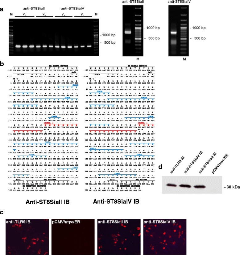
Background: Polysialic acid (polySia) is a carbohydrate modification of the neural cell adhesion molecule (NCAM), which is implicated in neural differentiation and plays an important role in tumor development and metastasis. Polysialylation of NCAM is mediated by two Golgi-resident polysialyltransferases (polyST) ST8SiaII and ST8SiaIV. Intracellular antibodies (intrabodies; IB) expressed inside the ER and retaining proteins passing the ER such as cell surface receptors or secretory proteins provide an efficient means of protein knockdown. To inhibit the function of ST8SiaII and ST8SiaIV specific ER IBs were generated starting from two corresponding hybridoma clones. Both IBs αST8SiaII-IB and αST8SiaIV-IB were constructed in the scFv format and their functions characterized in vitro and in vivo.

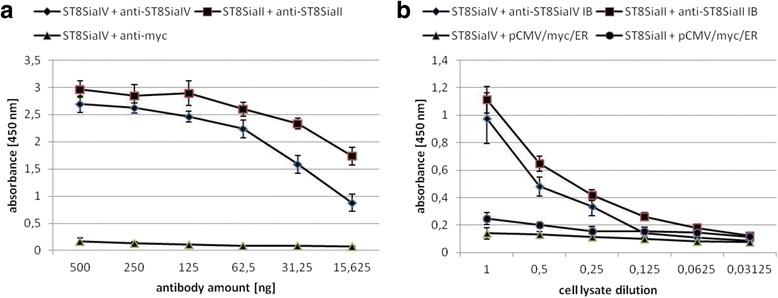
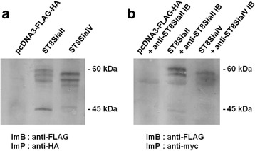
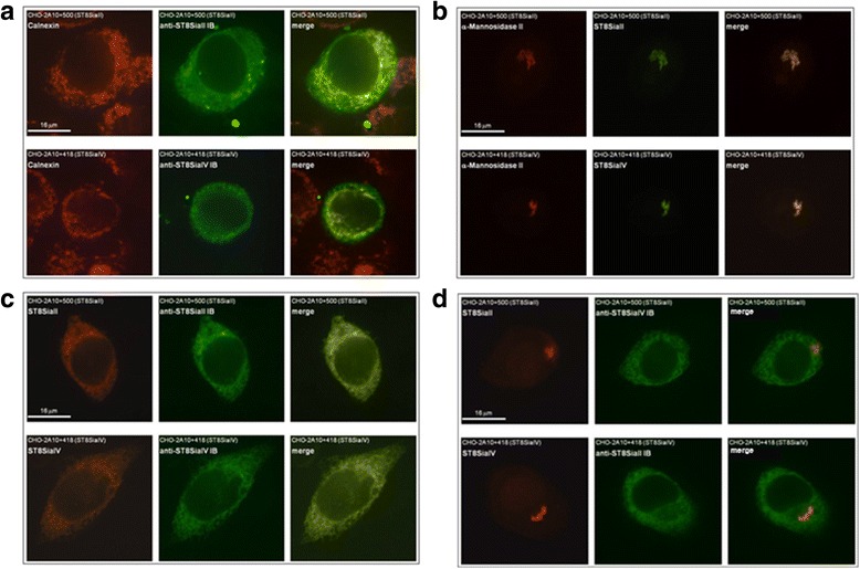
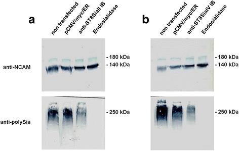
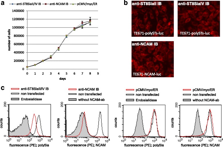
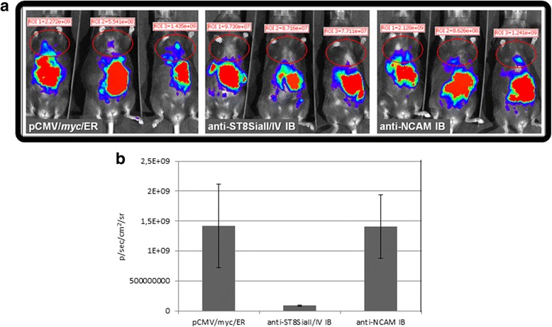
Results: IBs directed against the polySTs prevented the translocation of the enzymes from the ER to the Golgi-apparatus. Co-immunoprecipitation of ST8SiaII and ST8SiaIV with the corresponding IBs confirmed the intracellular interaction with their cognate antigens. In CHO cells overexpressing ST8SiaII and ST8SiaIV, respectively, the transfection with αST8SiaII-IB or αST8SiaIV-IB inhibited significantly the cell surface expression of polysialylated NCAM. Furthermore stable expression of ST8SiaII-IB, ST8SiaIV-IB and luciferase in the rhabdomyosarcoma cell line TE671 reduced cell surface expression of polySia and delayed tumor growth if cells were xenografted into C57BL/6 J RAG-2 mice.

Conclusion: Data obtained strongly indicate that αST8SiaII-IB and αST8SiaIV-IB are promising experimental tools to analyze the individual role of the two enzymes during brain development and during migration and proliferation of tumor cells.

Keywords: ER intrabodies; Neural cell adhesion molecule; Polysialic acid cell surface expression; Polysialyltransferases; Xenograft tumor mouse model.


Figures

Fig. 1
Fig. 1
Assembly and expression of ER IBs recognizing ST8SiaII and ST8SiaIV. a PCR amplification of the variable domains VH and VL of anti-ST8SiaII and anti-ST8SiaiIV scFv fragments (left side) and assembly of the corresponding scFvs (right side). 20 μl of the PCR products were analyzed on a 1% agarose gel (M = 100 bp ladder). b Primary sequence of anti-ST8SiaII and anti-ST8SiaIV intrabody. Shown are the coding (lower lane) and amino acid sequence (upper lane) of anti-ST8SiaII-IB and anti-ST8SiaIV-IB including the ER signal peptide, the myc epitope and the ER retention sequence. The complementary-determing regions (CDR1-CDR3) of the variable domains of the heavy and light chain are printed in blue letters. The synthetic linker, shown in red letters, localized between the VH and VL domains was introduced by assembly PCR. c Immunofluorescence analysis by microscopy of permeabilized and fixed HEK293 cells transiently transfected with IBs. Intracellular expression was stained in red. Positive control: anti-TLR9 IB, negative control: HEK 293 cells transiently transfected with the empty expression plasmid pCMV/myc/ER. d Immunoblot analysis, Expression of anti-ST8SiaII-IB and anti-ST8SiaIV-IB visualized with peroxidase labelled secondary antibody. Sample volume: 10 μl of 100 μl cell lysat from 106 cells transiently transfected for 48 h with the intrabody DNA in a 6-well microtiter plate
Fig. 2
Fig. 2
Binding of anti-ST8SiaII-IB and anti-ST8SiaIV-IB to their antigens in ELISA. a 50 ng purified ST8SiaII and ST8SiaIV in 50 μl 0.2 M sodium phosphate puffer was immobilized on MaxiSorb™ polystyrene assay plates (Nunc) as indicated. Serial dilutions of purified original anti-ST8SiaII and anti-ST8SiaIV mAbs 3167 and 3175, respectively, were applied in 100 μl PBS. Negative control: ST8SiaIV incubated with anti-myc antibody. b Serial dilutions of 100 μl cell lysates of 106 HEK293 cells transiently transfected with anti-ST8SiaII-IB expression plasmid or anti-ST8SiaIV-IB expression plasmid for 48 h in a 6 well microtiter plate were incubated in different serial dilutions in 100 μl PBS with immobilized purified ST8SiaII or ST8SiaIV. Negative controls: Cell lysates transfected with pCMV/myc/ER. Result of 3 independent experiments. Bars demonstrate standard deviation calculated from the mean values
Fig. 3
Fig. 3
Intracellular Binding of anti-ST8SiaII-IB and anti-ST8SiaIV-IB to their antigens. a control, immunoprecipitation of FLAG-HA tagged ST8SiaII and FLAG-HA tagged ST8SiaIV transiently transfected in 106 HEK293 cells for 48 h in a 6-well microtitre plate. After lysis in 100 μl lysisbuffer and immunoprecipitation the different glycosylated forms were analyzed by immunoblotting. Negative control: transfection with empty vector pcDNA3-FLAG-HA. b Co-IP of HEK 293 cells cotransfected with FLAG-HA tagged ST8SiaII and ST8SiaII-IB or FLAG-HA tagged ST8SiaIV and ST8SiaIV-IB. Negative control: Co-IP of HEK293 cells cotransfected with pcDNA3-FLAG-HA and anti-ST8SiaII-IB expressionplasmid. ImB: Immunoblot; ImP: Immunoprecipitation. Sample volume is 12 μl from total 25 μl immunoprecipitat. The experiment was done 2 times, shown is a representative example
Fig. 4
Fig. 4
Anti-ST8SiaII-IB and anti-ST8SiaIV-IB mediates retention of ST8SiaII and ST8SiaIV inside the ER. 106 recombinant CHO cells transiently transfected for 48 h with the corresponding intrabody DNA or unspecific intrabody DNA in a 6-well microtiter plate were analyzed by immunofluorescence. a Colocalisation of IBs with ER marker Calnexin. Recombinant CHO cells expressing ST8SiaII (CHO-2A10 + 500) were transfected with anti-ST8SiaII-IB expression plasmid (upper line) or CHO cells expressing ST8SiaIV (CHO-2A10 + 418) were transfected with anti-ST8SiaIV-IB expression plasmid (lower line). ER resident marker Calnexin was stained red. IBs were labelled green. b colocalisation of ST8SiaII and ST8SiaIV in non transfected CHO-2A 10 + 500 cells (upper line) and in non transfected CHO-2A10 + 418 cells (lower line) with Golgi marker α-mannose II. Golgi marker α-mannose II was stained red. Polysialyltransferases were labeled green. c ST8SiaII and anti-ST8SiaII-IB or ST8SiaIV and anti-ST8SiaIV-IB were stained in anti-ST8SiaII-IB or ST8SiaIV-IB transfected CHO-2A10 + 500 cells or CHO-2A10 + 418 cells. In this case the polysialyltransferases were stained red d negative controls: Staining of the polysialyltransferases and IBs in the CHO cells transfected with the unspecific IBs. The experiment was done 3 times. Shown is a characteristic staining
Fig. 5
Fig. 5
Reduced expression of polySia in recombinant CHO cells mediated by anti-ST8SiaII and anti-ST8SiaIV IBs. a Immunoblot analysis of expressed NCAM and polySia of NCAM in CHO-2A10 + 500-cells expressing ST8SiaII transfected with ST8SiaII-IB. Negative control: cells non transfected or transfected with pCMV/myc/ER. Positive control: cell lysat treated with endosialidase. b Immunoblot analysis of expressed NCAM and polySia of NCAM in CHO-2A10 + 418-cells expressing ST8SiaIV transfected with ST8SiaIV IB. The experiment was done 3 times. Shown is a characteristic example. Sample volume: 10 μl of 100 μl cell lysat from 106 cells grown for 48 h in a 6-well microtiter plate
Fig. 6
Fig. 6
PolySia cell surface expression is inhibited in TE671 rhabdomyosarcoma cells expressing α ST8 Sia II and α ST8 Sia IV intrabody. a Proliferation assay of 5 x 10⁴ TE671 cells stable expressing αST8SiaII-IB and αST8SiaIV-IB, anti-NCAM IB or empty vector cultured in wells of a 6-well microtiter plate over a time period of 8 days. The number of living cells was determined after staining with trypan blue using a Rosenthal-Fuchs counting chamber. Starting material was 5 × 10⁴ cells. The proliferation assay was done 3 times, bars demonstrate standard deviation calculated from the mean values. b Immunofluorescence analysis of anti-ST8SiaII-IB, anti-ST8SiaIV-IB and anti-NCAM intrabody expression. 106 recombinant CHO cells transiently transfected for 48 h with the anti-PolySTs intrabodies or anti-NCAM intrabody in a 6-well microtiter plate were analyzed by immunofluorescence. Anti-ST8SiaII, Anti-ST8SiaIV-IB and anti-NCAM intrabody were detected in red. c Flow cytometric detection of polySia and NCAM cell surface expression. Stable transfected cell lines are shown with red lines. Negative controls are non-transfected cells (grey line) and cells only incubated with the secondary antibody (grey area). In the case of detection of polySia negative controls are cells treated with endosialidase (grey area). 104 cells in 300 μl PBS containing 2% FCS and 10 μg/ml propidiumiodide were measured. The flow cytometric analysis was performed 4 times. Shown is characteristic cell surface staining of NCAM and polySia from one experiment
Fig. 7
Fig. 7
Inhibitory effect of anti-ST8SiaII-IB and anti-ST8SiaIV-IB on metastasis of rhabdyomasarcoma cells after 4 weeks of tumor cell injection in mice. a Detection of tumor cells by luminescence. 106 rhabdomyosarcoma cells in 100 μl PBS expressing anti-ST8SiaII-IB and anti-ST8SiaIV-IB or anti-NCAM-IB or as negative control tumor cells stable transfected with the empty vector were injected intraperitoneally into 3 C57BL/6 J RAG-2 mice at a time. Luminiscence was determined at week 4 using in vivo imaging systems (IVIS). Shown are the results as luminiscence signals (p/sec/cm2/sr). ROI: region of interest. The red circles shows the metastic tumor cells in the region of lung and liver. b Mean values of each group of mice with corresponding standard deviation. Tumor growth was tracked every week over a period of 6 weeks. Complete tumor tracking is shown in supplement data

Similar articles

Cited by

References

    1. Rougon G, Hobert O. New insights into the diversity and function of neuronal immunoglobulin superfamily molecules. Annu Rev Neurosci. 2003;26:207–238. doi: 10.1146/annurev.neuro.26.041002.131014. - DOI - PubMed
    1. Edelman GM, Crossin KL. Cell adhesion molecules: implications for a molecular histology. Annu Rev Biochem. 1991;60:155–190. doi: 10.1146/annurev.bi.60.070191.001103. - DOI - PubMed
    1. Rothbard JB, Brackenbury R, Cunningham BA, Edelman GM. Differences in the carbohydrate structures of neural cell-adhesion molecules from adult and embryonic chicken brains. J Biol Chem. 1982;257(18):11064–11069. - PubMed
    1. Nelson RW, Bates PA, Rutishauser U. Protein determinants for specific polysialylation of the neural cell adhesion molecule. J Biol Chem. 1995;270(29):17171–17179. doi: 10.1074/jbc.270.29.17171. - DOI - PubMed
    1. Finne J. Occurrence of unique polysialosyl carbohydrate units in glycoproteins of developing brain. J Biol Chem. 1982;257(20):11966–11970. - PubMed

MeSH terms