. 2017 Sep:110:11-18.
doi: 10.1016/j.freeradbiomed.2017.05.006. Epub 2017 May 9.

Cyclophosphamide and acrolein induced oxidative stress leading to deterioration of metaphase II mouse oocyte quality

Affiliations

Cyclophosphamide and acrolein induced oxidative stress leading to deterioration of metaphase II mouse oocyte quality

Roohi Jeelani et al. Free Radic Biol Med. 2017 Sep.

Abstract

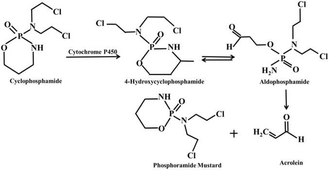
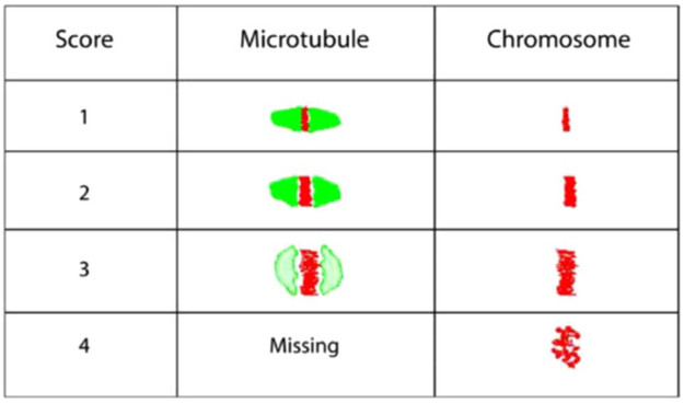
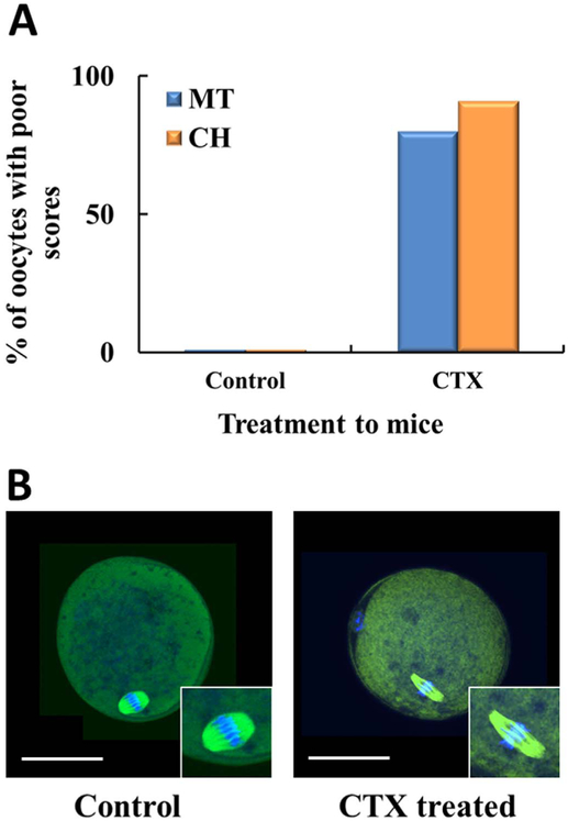
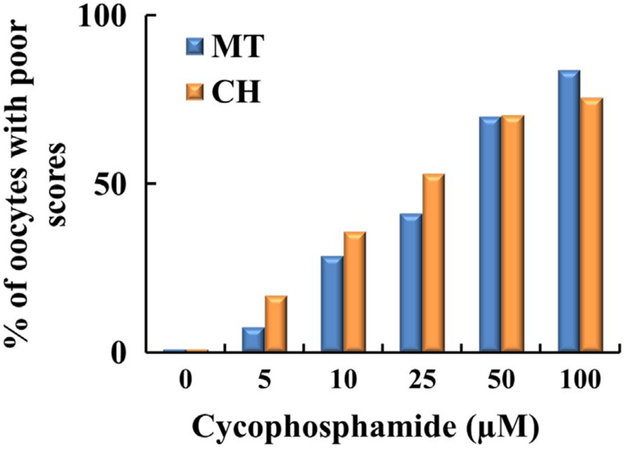
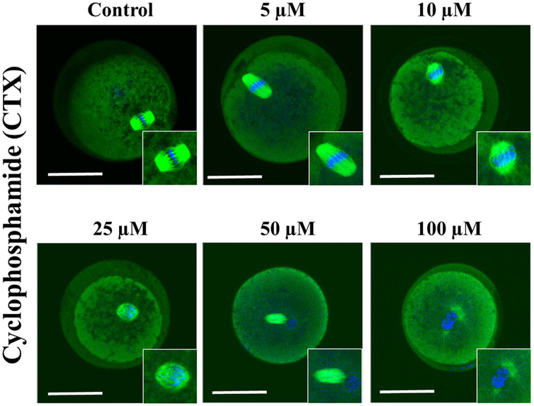
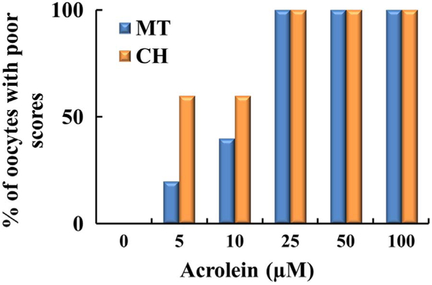
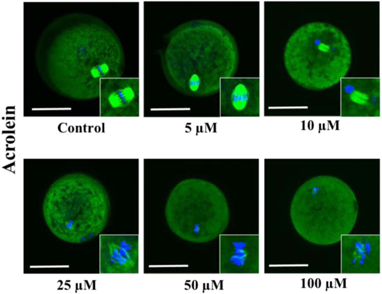
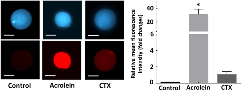
Cyclophosphamide (CTX) is a chemotherapeutic agent widely used to treat ovarian, breast, and hematological cancers as well as autoimmune disorders. Such chemotherapy is associated with reproductive failure and premature ovarian insufficiency. The mechanism by which CTX and/or its main metabolite, acrolein, affect female fertility remains unclear, but it is thought to be caused by an overproduction of reactive oxygen species (ROS). Here, we investigated the effect of CTX on metaphase II mouse oocytes obtained from treated animals (120mg/kg, 24h of single treatment), and oocytes directly exposed to increasing concentrations of CTX and acrolein (n=480; 0, 5, 10, 25, 50, and 100μM) with and without cumulus cells (CCs) for 45min which correlates to the time of maximum peak plasma concentrations after administration. Oocytes were fixed and subjected to indirect immunofluorescence and were scored based on microtubule spindle structure (MT) and chromosomal alignment (CH). Generation of ROS was evaluated using the Cellular Reactive Oxygen Species Detection Assay Kit. Deterioration of oocyte quality was noted when oocytes were obtained from CTX treated mice along with CTX and acrolein treated oocytes in a dose-dependent manner as shown by an increase in poor scores. Acrolein had an impact at a significantly lower level as compared to CTX, plateau at 10μM versus 50μM, respectively. These variation is are associated with the higher amount of ROS generated with acrolein exposure as compared to CTX (p<0.05). Utilization of antioxidant therapy and acrolein scavengers may mitigate the damaging effects of these compounds and help women undergoing such treatment.

Keywords: Acrolein; Chemotherapy; Chromosome alignment; Cyclophosphamide; Follicle dysfunction; Infertility; Meiotic spindle; Oocyte quality; Oxidative stress.


Figures

Fig. 1.
Fig. 1.
Mechanism of cyclophosphamide breakdown through the cytochrome P450 system to two stable toxic metabolites: Phosphoramide mustard and acrolein through the formation of unstable transient Intermediates: 4-hydroxycyclophosphamide and aldophosphamide.
Fig. 2.
Fig. 2.
Oocyte scoring system based on microtubule and chromosomes. The oocytes are scored from a score of 1–4. A score of 1 and 2 are considered good quality oocytes as the microtubules are well organized, and the chromosome is dense and tightly aligned. As the scores progress to 3 and 4, which are considered poor quality scores you see a ballooning effect of the microtubule and eventual loss and the chromosome starts dispersing away from the midline and scatters.
Fig. 3.
Fig. 3.
Intraperitoneal injection of cyclophosphamide in mice deteriorates oocyte quality. Two separate groups of female mice with a total of three mice in each group. Group 1 cyclophosphamide (treatment group) intraperitoneal injection (IP) and group 2 (control group) with Phosphate Buffer IP. All females from groups 1 & 2 were sacrificed and the oviducts were collected. The oocytes were collected, placed in fixative, primary and secondary antibody, DAPI and scored. A) There was a decline in oocyte quality based on MT and CH in the treatment group. The oocyte score is an average of three scores. B) Confocal microscopy to assess microtubules and spindle of CTX treated mice. Representative images of the control oocyte with normal spindle and chromosomal (CH) alignment as compared to the CTX treated mouse with a thin long spindle and outward movement of the chromosome from the equatorial plate.
Fig. 4.
Fig. 4.
Plot demonstrating percentage of oocytes with poor scores based on increasing concentration of cyclophosphamide. Oocytes (n = 240) were incubated with increasing concentrations of cyclophosphamide (0–100 μM). As the concentration increased, there was a progressive increase in poor scores based on MT, the blue column and the chromosomes which are represented by the orange column. Each treatment is the average of at least three independent experiments.
Fig. 5.
Fig. 5.
Confocal microscopy to assess microtubules and spindle in the presence of increasing concentration of cyclophosphamide. Starting with the control oocyte with normal spindle and chromosomal (CH) alignment, then as the concentration increased progressively altered spindle and CH alignment with the maximum deterioration of oocyte quality noted at 100 μM. Scale bars: 50 μm. Results depict observations made after three experiments. (For interpretation of the references to color in this figure, the reader is referred to the web version of this article).
Fig. 6.
Fig. 6.
Plot demonstrating percentage of oocytes with poor scores based on increasing concentration of acrolein. Oocytes (n = 240) were incubated with increasing concentrations of acrolein (0–100 μM). As the concentration is increased of acrolein, there was a progressive decline in oocyte quality based on MT and CH. Each treatment is the average of at least three independent experiments.
Fig. 7.
Fig. 7.
Confocal microscopy to assess microtubules and spindle in the presence of increasing concentration of acrolein. Starting with the control oocyte with normal spindle and chromosomal (CH) alignment, then as the concentration increased progressively altered spindle and CH alignment with significant deterioration noted at even 10 μM of acrolein. Scale bars: 50 μm. Results depict observations made after three experiments. (For interpretation of the references to color in this figure, the reader is referred to the web version of this article).
Fig. 8.
Fig. 8.
Verification of generation of damage to the oocytes was mediated through the production of reactive oxygen species. Oocytes without CCs (n = 60) were exposed to cyclophosphamide and acrolein for 45-min incubation to determine intracellular ROS generation. The fluorescence intensity was estimated as relative fluorescence unit (RFU) and plotted as a bar graph to present the fold changes in ROS production upon treatment relative to untreated control oocytes. Error bars indicate ± relative standard error of mean (SEM). * p < 0.05 vs. controls. Inset presents representative images of control and oocytes exposed to each compound for 45 min. Scale bars: 50 μm. Images shown are from a typical experiment performed at least three times.

References

    1. Guida M, et al., Reproductive issues in patients undergoing hematopoietic stem cell transplantation: an update, J. Ovarian Res 9 (1) (2016) 72. - PMC - PubMed
    1. Morgan S, et al., How do chemotherapeutic agents damage the ovary? Hum. Reprod. Update 18 (5) (2012) 525–535. - PubMed
    1. Blumenfeld Z, Chemotherapy and fertility, Best. Pract. Res. Clin. Obstet. Gynaecol 26 (3) (2012) 379–390. - PubMed
    1. Garrido-Colino C, et al., Current situation on fertility preservation in cancer patients in Spain: level of knowledge, information, and professional involvement, An. Pediatr (2016). - PubMed
    1. Nieman CL, et al., Cancer survivors and infertility: a review of a new problem and novel answers, J. Support Oncol 4 (4) (2006) 171–178. - PubMed

MeSH terms

LinkOut - more resources