

Case Reports
doi: 10.1136/bcr-2016-219172.
Angioleiomyoma of the upper lip
Affiliations
- PMID: 28500121
- PMCID: PMC5612575
- DOI: 10.1136/bcr-2016-219172
Item in Clipboard
Case Reports
Angioleiomyoma of the upper lip
BMJ Case Rep.
.
Abstract
This report describes a case of labial angioleiomyoma in a 52-year-old woman. The patient had noticed a slow-growing painless isolated mass in her upper lip for 6 months. The mass was surgically excised, and pathological examination was consistent with angioleiomyoma. Surgical excision was curative, and there was no recurrence at 12-month follow-up.
Keywords: Dentistry and oral medicine; Ear, nose and throat/otolaryngology; Mouth.
© BMJ Publishing Group Ltd (unless otherwise stated in the text of the article) 2017. All rights reserved. No commercial use is permitted unless otherwise expressly granted.
Conflict of interest statement
Competing interests: None declared.
Figures
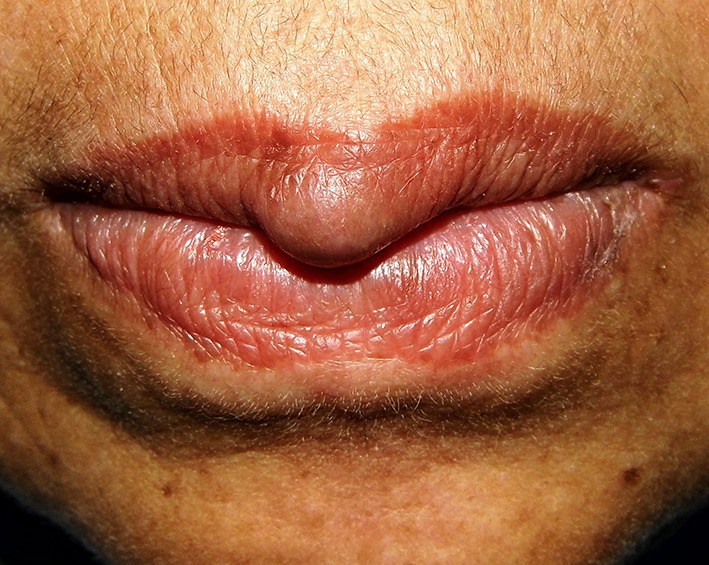
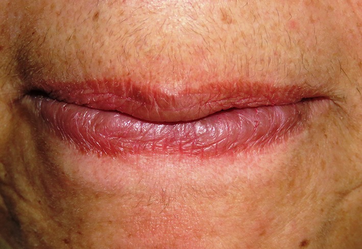
Clinical examination revealed the presence of a pea-sized mass occupying the central zone of the upper lip. The mass was firm, well-circumscribed, non-tender to palpation, covered by normal vermilion mucosa, and did not blanch with pressure.
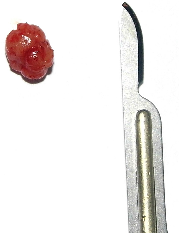
Macroscopic examination revealed a small mass which was well-circumscribed, light-brown in colour, firm in consistency, oval in shape and having a smooth surface.
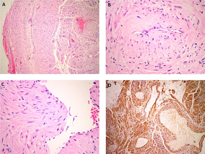
(A) Microscopic examination (H&E) revealed that the mass was well-circumscribed and composed of vascular channels. (B) The vascular channels were surrounded by fascicles of concentrically arranged spindle cells. (C) Tumour cells exhibited eosinophilic cytoplasm and oval nuclei showing no mitotic figures and a minimal degree of pleomorphism. (D) Immunohistochemical examination (SMA) revealed that the spindle cells were strongly positive for smooth muscle actin (SMA). In addition, the walls of vascular spaces were also positive for SMA.
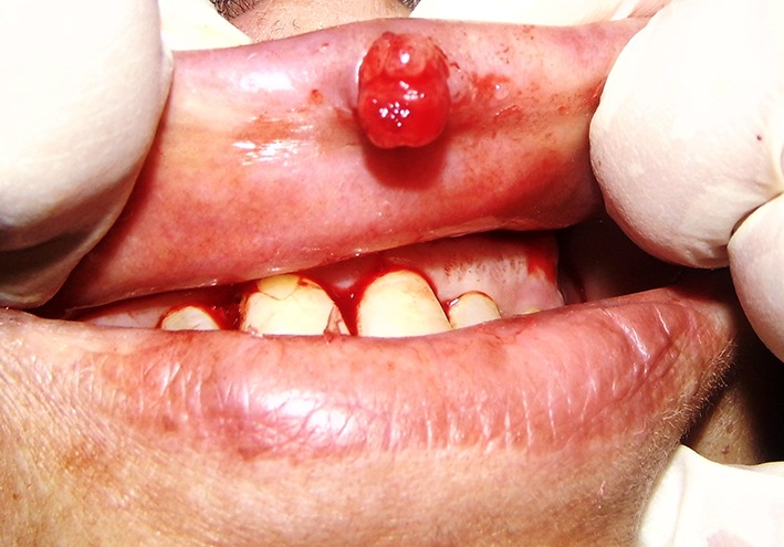
Complete surgical excision was performed under local anaesthesia. The mass was not adherent to the surrounding tissues and was removed in one piece.
At 12-month follow-up, there was no recurrence, and the area had healed without complications.
References
-
- Enzinger FM, Lattes R, Torloni H. Histological typing of soft tissue tumours. Geneva: World Health Organization, 1969:30–1.
-
- Morimoto N. Angiomyoma. A clinicopathological study. Med J Kagoshima Univ 1973;24:663–83.
Publication types
MeSH terms
LinkOut - more resources
Full Text Sources
Other Literature Sources