. 2017 May 12;7(1):1866.
doi: 10.1038/s41598-017-02049-9.

Microgravity induces inhibition of osteoblastic differentiation and mineralization through abrogating primary cilia

Affiliations

Microgravity induces inhibition of osteoblastic differentiation and mineralization through abrogating primary cilia

Wengui Shi et al. Sci Rep. .

Abstract

It is well documented that microgravity in space environment leads to bone loss in astronauts. These physiological changes have also been validated by human and animal studies and modeled in cell-based analogs. However, the underlying mechanisms are elusive. In the current study, we identified a novel phenomenon that primary cilia (key sensors and functioning organelles) of rat calvarial osteoblasts (ROBs) gradually shrank and disappeared almost completely after exposure to simulated microgravity generated by a random positioning machine (RPM). Along with the abrogation of primary cilia, the differentiation, maturation and mineralization of ROBs were inhibited. We also found that the disappearance of primary cilia was prevented by treating ROBs with cytochalasin D, but not with LiCl or dynein light chain Tctex-type 1 (Dynlt1) siRNA. The repression of the differentiation, maturation and mineralization of ROBs was effectively offset by cytochalasin D treatment in microgravity conditions. Blocking ciliogenesis using intraflagellar transport protein 88 (IFT88) siRNA knockdown inhibited the ability of cytochalasin D to counteract this reduction of osteogenesis. These results indicate that the abrogation of primary cilia may be responsible for the microgravity's inhibition on osteogenesis. Reconstruction of primary cilia may become a potential strategy against bone loss induced by microgravity.


Conflict of interest statement

The authors declare that they have no competing interests.

Figures

Figure 1
Figure 1
The random positioning machine (RPM) used in this study to generate microgravity for cultured cells. (A) The schematic view of the desktop RPM. (B) A photo of the control console operating the outer and inner frames of the RPM to rotate the cell culture vessel separately and randomly at 3-dimension in the range of 0–10 rpm. (C) A photo showing the RPM placed in a cell incubator. Six 50 ml-flasks with cultured ROBs sealed by aerated hydrophobic membrane were fixed on the cell culture vessel of the RPM.
Figure 2
Figure 2
Simulated microgravity significantly abrogates the primary cilia of rat calvarial osteoblasts (ROBs). (A) The immunofluorescence image of ROBs with or without random positioning machine (RPM) microgravity treatment. Primary cilia were stained by acetylated α-tubulin (green), and the nuclei were stained with DAPI (blue). (B) The percentage of ciliolated cells with or without RPM treatment. (C) Means (red bars) and individual measurements of cilium lengths with or without RPM treatment. (D) The protein expression levels of acetylated α-tubulin in ROBs with or without RPM treatment. NG: normal ground, SMG: simulated microgravity. Full-length Western blots are presented in Supplementary Figure 1. Each experiment was conducted at least three times independently. *P < 0.05 or **P < 0.01 vs NG group or 0 h group.
Figure 3
Figure 3
Simulated microgravity inhibits the osteogenic differentiation of rat calvarial osteoblasts (ROBs). (A) The alkaline phosphatase (ALP) activities after ROBs were exposed to simulated microgravity (SMG) and normal ground (NG) for different time periods. (B) Representative Western blots detecting the expression levels of osteogenesis markers, COL-I, BMP-2 and Runx-2 (with GAPDH as a control) in ROBs at different time points after random positioning machine (RPM) exposure. (C) Relative protein levels of COL-I, BMP-2 and Runx-2. NG: normal ground, SMG: simulated microgravity. Full-length Western blots are presented in Supplementary Figure 2. Each experiment was conducted at least three times independently. *P < 0.05 or **P < 0.01 vs NG group or 0 h group.
Figure 4
Figure 4
Simulated microgravity reduces the maturation and mineralization of rat calvarial osteoblasts (ROBs). (A) Representative image of the alkaline phosphatase (ALP) stained CFU-FALP colonies after 8 days of random positioning machine (RPM) microgravity treatment. (B) Representative image of the stained mineralized nodules after 12 days of RPM treatment. (C) Enlarged image of the CFU-FALP colonies under a phase contrast microscope. (D) Enlarged image of the mineralized nodules under a phase contrast microscope. (E and F) The numbers and areas of the CFU-FALP . (G and H) The numbers and areas of the mineralized nodules. NG: normal ground, SMG: simulated microgravity. Each experiment was conducted at least three times independently. *P < 0.05 or **P < 0.01 vs NG group.
Figure 5
Figure 5
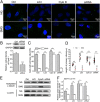
Elongation of cilia prevents the abrogation of primary cilia induced by simulated microgravity. (A) Immunofluorescence image of rat calvarial osteoblasts (ROBs) exposed to simulated microgravity (SMG) or normal ground (NG) for 24 h after being treated with 5 mM LiCl, 0.1 μM cytochalasin D (Cyto D) or dynein light chain Tctex-type 1 (Dynlt1) siRNA. Primary cilia were stained with acetylated α-tubulin (green), and DNA were stained with DAPI (blue). (B) Treatment with Dynlt1 siRNA resulted in significant decreases in Dynlt1 expression at the protein levels. (C) Quantification of the percentage of cells with a cilium in each group. (D) Means (red bars) and individual measurements of cilium lengths in each group. (E) The protein expression levels of acetylated α-tubulin. Full-length Western blots are presented in Supplementary Figure 3 and Figure 4. Each experiment was conducted at least three times independently. *P < 0.05 or **P < 0.01 vs NG group of each treatment; & P < 0.05 or && P < 0.01 vs Ctrl of NG group; △△ P < 0.01 vs Ctrl of SMG groups.
Figure 6
Figure 6
Elongation of cilia promotes the osteoblastic differentiation of rat calvarial osteoblasts (ROBs) in microgravity condition. (A) Alkaline phosphatase (ALP) activities of ROBs in Ctrl group, LiCl group, cytochalasin D (Cyto D) group or dynein light chain Tctex-type 1 (Dynlt1) siRNA group with or without random positioning machine (RPM) microgravity exposure. (B) Representative Western blot detecting expression of osteogenesis markers COL-I, BMP-2 and Runx-2 (with GAPDH as a control) in each group. (C) The protein levels of COL-I, BMP-2 and Runx-2 in each group. NG: normal ground, SMG: simulated microgravity. Full-length Western blots are presented in Supplementary Figure 5. Each experiment was conducted at least three times independently. *P < 0.05 or **P < 0.01 vs NG group of each treatment; △△ P < 0.01 vs Ctrl of SMG groups.
Figure 7
Figure 7
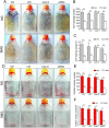
Elongation of cilia promotes the maturation and mineralization of rat calvarial osteoblasts (ROBs) in microgravity condition. (A) Representative images of alkaline phosphatase (ALP) stained CFU-FALP colonies after 8 days in Ctrl group, LiCl group, cytochalasin D (Cyto D) group or dynein light chain Tctex-type 1 (Dynlt1) siRNA group with or without random positioning machine (RPM) microgravity exposure. (B and C) The numbers and areas of CFU-FALP in each group. (D) Representative image of stained mineralized nodules after 12 days in each group. (E and F) The numbers and areas of mineralized nodules in each group. NG: normal ground, SMG: simulated microgravity. Each experiment was conducted at least three times independently. *P < 0.05 or **P < 0.01 vs NG group of each treatment; △△ P < 0.01 vs Ctrl of SMG groups.
Figure 8
Figure 8
Intraflagellar transport protein 88 (IFT88) siRNA knockdown inhibits the ability of cytochalasin D (Cyto D) to counteract the reduction of osteogenesis. (A) Immunofluorescence image of rat calvarial osteoblasts (ROBs) treated with or without IFT88 siRNA and 0.1 μM Cyto D. Primary cilia were stained with acetylated α-tubulin (green), and DNA were stained with DAPI (blue). (B) Treatment with IFT88 siRNA resulted in significant decreases in IFT88 expression at the protein levels. (C) Quantification of the percentage of cells with a cilium in each group. (D) Representative Western blot detecting expression of osteogenesis markers COL-I, BMP-2 and Runx-2 (with GAPDH as a control) in each group. (E-G) The protein levels of COL-I, BMP-2 and Runx-2 in each group. NG: normal ground, SMG: simulated microgravity. Full-length Western blots are presented in Supplementary Figures 6 and 7. Each experiment was conducted at least three times independently. **P < 0.01 vs NG group of each treatment; △△ P < 0.01 vs Ctrl of SMG groups.

Similar articles

Cited by

References

    1. Loomer PM. The impact of microgravity on bone metabolism in vitro and in vivo. Crit. Rev. Oral. Biol. Med. 2001;12:252–261. doi: 10.1177/10454411010120030401. - DOI - PubMed
    1. Kramer LA, Sargsyan AE, Hasan KM, Polk JD, Hamilton DR. Orbital and intracranial effects of microgravity: findings at 3-T MR imaging. Radiology. 2012;263:819–827. doi: 10.1148/radiol.12111986. - DOI - PubMed
    1. Leach CS. Biochemical and hematologic changes after short-term space flight. Microgravity Q. 1992;2:69–75. - PubMed
    1. Mccarthy ID. Fluid shifts due to microgravity and their effects on bone: a review of current knowledge. Ann. Biomed. Eng. 2005;33:95–103. doi: 10.1007/s10439-005-8967-6. - DOI - PubMed
    1. Bucaro MA, et al. Bone cell survival in microgravity: evidence that modeled microgravity increases osteoblast sensitivity to apoptogens. Ann. NY. Acad. Sci. 2004;1027:64–73. doi: 10.1196/annals.1324.007. - DOI - PubMed

Publication types

LinkOut - more resources