. 2017 May 12;7(1):1864.
doi: 10.1038/s41598-017-02041-3.

Examining the efficacy of intravenous administration of predatory bacteria in rats

Affiliations

Examining the efficacy of intravenous administration of predatory bacteria in rats

Kenneth Shatzkes et al. Sci Rep. .

Abstract

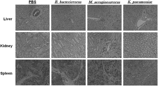
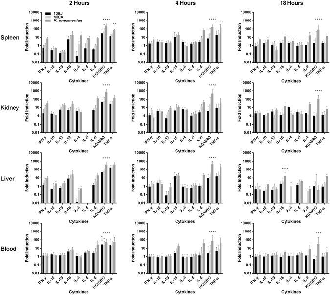
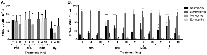
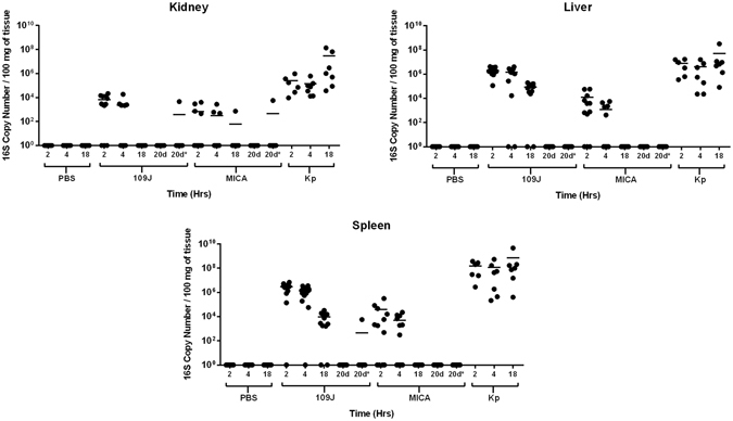
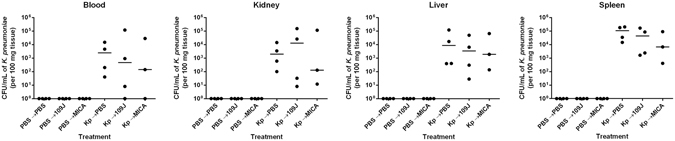
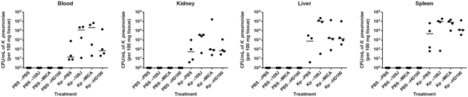
The proteobacteria Bdellovibrio bacteriovorus and Micavibrio aeruginosavorus are obligate predators of Gram-negative bacteria, and have been proposed to be used to treat multidrug-resistant bacterial infections. The ability of predatory bacteria to reduce bacterial burden in vivo within the lungs of rats has been demonstrated, but it was unknown if predatory bacteria can attenuate systemic bacterial burden administered intravenously. In this study, we first assessed the safety of intravenous inoculation of predatory bacteria in rats. No rat morbidity or adverse histopathology of various organs due to predatory bacteria administration was observed. An increase in proinflammatory cytokines (TNFα and KC/GRO) was observed at two hours post-inoculation; however, cytokines returned to baseline levels by 18 hours. Furthermore, bacterial dissemination analysis demonstrated that predatory bacteria were efficiently cleared from the host by 20 days post-injection. To determine whether predatory bacteria could reduce bacterial burden in vivo, Klebsiella pneumoniae was injected into the tail veins of rats and followed with multiple doses of predatory bacteria over 16 or 24 hours. Predatory bacteria were unable to significantly reduce K. pneumoniae burden in the blood or prevent dissemination to other organs. The results suggest that predatory bacteria may not be effective for treatment of acute blood infections.


Conflict of interest statement

The authors declare that they have no competing interests.

Figures

Figure 1
Figure 1
Histological examination of rat organs after intravenous inoculation of predatory bacteria. SD rats were intravenously inoculated (through tail vein injection) with PBS, B. bacteriovorus, M. aeruginosavorus, or K. pneumoniae. Histological examination of harvested rat livers, kidneys, and spleens exposed to B. bacteriovorus and M. aeruginosavorus revealed no abnormal pathology compared to rats treated with PBS. All images are representative micrographs that were taken at 18 hours post-inoculation and at X40 total magnification.
Figure 2
Figure 2
Inflammatory protein profile within rat blood and organs in response to intravenous inoculation of predatory bacteria. ELISA analysis of IL-1β, IL-4, IL-5, IL-6, IL-10, IL-13, CXCL-1/KC, IFNγ, and TNF in response to tail vein injection with predatory bacteria relative to PBS control was performed. Rats were injected with PBS, B. bacteriovorus 109J, or M. aeruginosavorus (and also K. pneumoniae [Kp] as a control). Inflammatory proteins were assessed within the spleen, liver, kidney and blood at two, four, and 18 hours post-inoculation. Twelve rats per treatment group (seven for K. pneumoniae) were used at each time point. Data are combined from two independent experiments. Data represent means ± standard errors of the means. Significant differences between treatment groups and respective control were determined using ANOVA (*P < 0.05; **P < 0.01; ***P < 0.001; ****P < 0.0001).
Figure 3
Figure 3
Inflammatory cell response to intravenous injection of predatory bacteria. In order to profile the (A) total white blood cell counts and (B) inflammatory cell response in the blood due to predatory bacteria, rats were injected through the tail vein with PBS, B. bacteriovorus 109J, M. aeruginosavorus, or K. pneumoniae (Kp). Blood samples were assessed at two, four, and 18 hours post-injection. Data represent means ± standard errors of the means. Significant differences between treatment groups and respective PBS control were determined using ANOVA.
Figure 4
Figure 4
Predatory bacterial dissemination within host. qPCR detection of predatory bacteria within the host was performed. The kidneys, livers, and spleens were probed for B. bacteriovorus 109J, or M. aeruginosavorus (MICA), and K. pneumoniae at two, four, and 18 hours post-injection. Twelve rats per treatment group (seven for K. pneumoniae) were analyzed at each time point. Each data point represents a single rat’s respective bacterial load. Horizontal lines represent the mean of the results from each treatment set. Data are combined from the results of two independent experiments.
Figure 5
Figure 5
K. pneumoniae bacterial burden within rat blood and organs after treatment scheme #1 with predatory bacteria. K. pneumoniae (or PBS for control groups) was initially introduced into rats via intravenous inoculation. Animals were then treated via tail vein injection with PBS, B. bacteriovorus 109J or M. aeruginosavorus (MICA) at 30 minutes, 6, 12, and 18 hours post-injection. At 24 hours, blood was collected and kidneys, livers, and spleens were harvested, homogenized, and plated on MacConkey agar plates to recover K. pneumoniae CFUs. Four rats per treatment group were used at each time point. Each data point represents a single rat’s respective bacterial load. Horizontal lines represent the median of the results from each treatment set. Significant differences between treatment groups and respective control were performed using the Mann-Whitney test.
Figure 6
Figure 6
K. pneumoniae bacterial burden within rat blood and organs after treatment scheme #2 with predatory bacteria. K. pneumoniae (or PBS for control groups) was initially introduced into rats via intravenous inoculation. Animals were then treated via tail vein injection with PBS, B. bacteriovorus 109J, HD100, or M. aeruginosavorus (MICA) at 30 minutes, 5, 9.5, and 14 hours post-injection. At 16 hours, blood was collected and kidneys, livers, and spleens were harvested, homogenized, and plated on MacConkey agar plates to recover K. pneumoniae CFUs. Four rats per treatment group were used at each time point. Each data point represents a single rat’s respective bacterial load. Horizontal lines represent the median of the results from each treatment set. Significant differences between treatment groups and respective control were performed using the Mann-Whitney test.

References

    1. Stolp H, Starr MP. Bdellovibrio bacteriovorus gen. et sp. n., a predatory, ectoparasitic, and bacteriolytic microorganism. Antonie Van Leeuwenhoek. 1963;29:217–248. doi: 10.1007/BF02046064. - DOI - PubMed
    1. Lambina VA, Afinogenova AV, Romay Penobad Z, Konovalova SM, Andreev LV. [New species of exoparasitic bacteria of the genus Micavibrio infecting gram-positive bacteria] Mikrobiologiia. 1983;52:777–780. - PubMed
    1. Chanyi RM, Koval SF. Role of type IV pili in predation by Bdellovibrio bacteriovorus. PLoS One. 2014;9:e113404. doi: 10.1371/journal.pone.0113404. - DOI - PMC - PubMed
    1. Evans KJ, Lambert C, Sockett RE. Predation by Bdellovibrio bacteriovorus HD100 requires type IV pili. J. Bacteriol. 2007;189:4850–4859. doi: 10.1128/JB.01942-06. - DOI - PMC - PubMed
    1. Mahmoud KK, Koval SF. Characterization of type IV pili in the life cycle of the predator bacterium Bdellovibrio. Microbiology. 2010;156:1040–1051. doi: 10.1099/mic.0.036137-0. - DOI - PubMed

Publication types