

The effect of repetitive flexion and extension fatigue loading on the young porcine lumbar spine, a feasibility study of MRI and histological analyses
- PMID: 28500483
- PMCID: PMC5429315
- DOI: 10.1186/s40634-017-0091-7
The effect of repetitive flexion and extension fatigue loading on the young porcine lumbar spine, a feasibility study of MRI and histological analyses
Abstract
Background: The biomechanical mechanisms of failure of FSUs have been studied but the correlation of repetitive flexion and extension loadings to the initial phase of fatigue in young FSUs are still not known. The purpose of the study was to examine the fatigue results of low magnitude repetitive flexion and extension loading on porcine lumbar Functional Spinal Units (FSUs) with Magnetic Resonance Imaging (MRI) and histology.
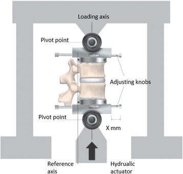
Methods: Eight FSUs were subject to repetitive pivot flexion and eight to extension loading by a protocol of 20 000 cycles at 1 Hz with a load of 700 N. All loaded FSUs (N = 16) were examined with MRI and histology post loading. Three FSUs were examined with MRI as controls. Further three FSUs were non loaded histology controls.
Results: Fifteen (94%) of the loaded FSUs have decreased MRI signal in the growth zone of the superior vertebra and 12 (75%) in the inferior vertebrae. Fourteen (88%) FSUs have increased signal in the superior vertebral body. Fourteen (88%) FSUs have a reduced signal in all or any endplate. The histology morphometry displayed that the unstained parts of the epiphyseal growth zone were larger among the loaded FSUs (mean 29% vs 4%) and that the chondrocytes in the endplate and growth zones had abnormal structure and deformed extracellular matrix.
Conclusion: Repetitive loading of young porcine FSUs in both extension and flexion causes concurrent MRI and histological changes in the growth zones and endplates, which could be a first sign of fatigue and an explanation for the disc, apophyseal and growth zone injuries seen among adolescent athletes.
Keywords: Animal experimentation; End plate histology; Fatigue; Growth zone injury; In vitro; Intervertebral disc; MRI; Repetitive loading; Spine.
Figures


Similar articles
-
Fracture patterns of the adolescent porcine spine: an experimental loading study in bending-compression.Spine (Phila Pa 1976). 2005 Jan 1;30(1):75-82. doi: 10.1097/00007632-200501010-00014. Spine (Phila Pa 1976). 2005. PMID: 15626985
-
Vertebral fractures and separations of endplates after traumatic loading of adolescent porcine spines with experimentally-induced disc degeneration.Clin Biomech (Bristol). 2005 Dec;20(10):1046-54. doi: 10.1016/j.clinbiomech.2005.06.014. Epub 2005 Aug 15. Clin Biomech (Bristol). 2005. PMID: 16102879
-
Cyclical loading causes injury in and around the porcine proximal femoral physeal plate: proposed cause of the development of cam deformity in young athletes.J Exp Orthop. 2015 Dec;2(1):6. doi: 10.1186/s40634-015-0022-4. Epub 2015 Mar 8. J Exp Orthop. 2015. PMID: 26914874 Free PMC article.
-
Needle puncture in rabbit functional spinal units alters rotational biomechanics.J Spinal Disord Tech. 2015 Apr;28(3):E146-53. doi: 10.1097/BSD.0000000000000196. J Spinal Disord Tech. 2015. PMID: 25370985 Free PMC article.
-
Intervertebral disc herniation: studies on a porcine model exposed to highly repetitive flexion/extension motion with compressive force.Clin Biomech (Bristol). 2001 Jan;16(1):28-37. doi: 10.1016/s0268-0033(00)00063-2. Clin Biomech (Bristol). 2001. PMID: 11114441
Cited by
-
No Significant Change in MRI Abnormalities or Back Pain Prevalence in the Thoraco-Lumbar Spine of Young Elite Skiers Over a 2-Year Follow-Up.Open Access J Sports Med. 2022 Aug 18;13:69-76. doi: 10.2147/OAJSM.S366548. eCollection 2022. Open Access J Sports Med. 2022. PMID: 36003328 Free PMC article.
-
Is There a Relationship Between Workload and Occurrence of Back Pain and Back Injuries in Athletes?Front Physiol. 2020 Jul 24;11:894. doi: 10.3389/fphys.2020.00894. eCollection 2020. Front Physiol. 2020. PMID: 32792989 Free PMC article. Review.
-
Low occurrence of MRI spinal changes in elite climbing athletes; a cross-sectional study.BMC Sports Sci Med Rehabil. 2023 Mar 9;15(1):29. doi: 10.1186/s13102-023-00637-z. BMC Sports Sci Med Rehabil. 2023. PMID: 36895033 Free PMC article.
-
MRI-detected spinal disc degenerative changes in athletes participating in the Rio de Janeiro 2016 Summer Olympics games.BMC Musculoskelet Disord. 2020 Jan 20;21(1):45. doi: 10.1186/s12891-020-3057-3. BMC Musculoskelet Disord. 2020. PMID: 31959161 Free PMC article.
-
Effects of Competition Level on the Prevalence and Incidence of Lumbar Disk Degeneration in Japanese Collegiate Gymnasts.Orthop J Sports Med. 2022 Nov 15;10(11):23259671221119439. doi: 10.1177/23259671221119439. eCollection 2022 Nov. Orthop J Sports Med. 2022. PMID: 36419478 Free PMC article.
References
LinkOut - more resources
Full Text Sources
Other Literature Sources
