Review
. 2017 Jun;8(3):357-363.
doi: 10.1007/s13244-017-0555-4. Epub 2017 May 12.

Tips and tricks for a safe and effective image-guided percutaneous renal tumour ablation

Affiliations
Review

Tips and tricks for a safe and effective image-guided percutaneous renal tumour ablation

Giovanni Mauri et al. Insights Imaging. 2017 Jun.

Abstract

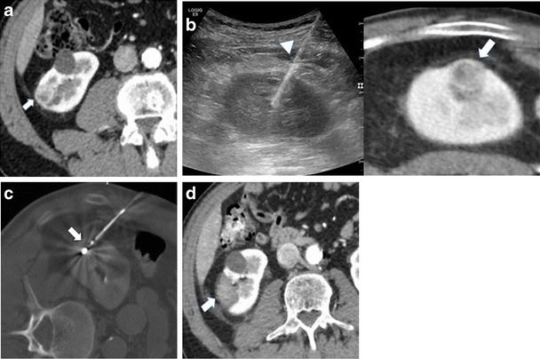
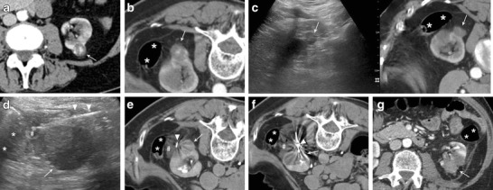
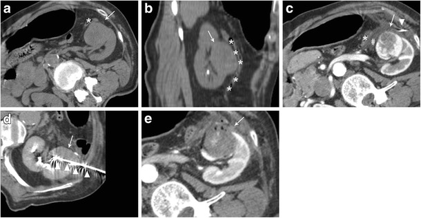
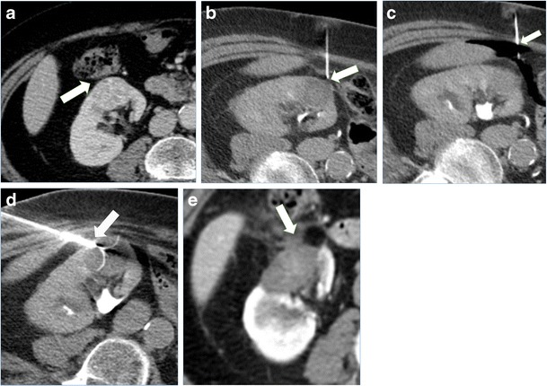
Image-guide thermal ablations are nowadays increasingly used to provide a minimally invasive treatment to patients with renal tumours, with reported good clinical results and low complications rate. Different ablative techniques can be applied, each with some advantages and disadvantages according to the clinical situation. Moreover, percutaneous ablation of renal tumours might be complex in cases where there is limited access for image guidance or a close proximity to critical structures, which can be unintentionally injured during treatment. In the present paper we offer an overview of the most commonly used ablative techniques and of the most important manoeuvres that can be applied to enhance the safety and effectiveness of percutaneous image-guided renal ablation. Emphasis is given to the different technical aspects of cryoablation, radiofrequency ablation, and microwave ablation, on the ideal operating room setting, optimal image guidance, application of fusion imaging and virtual navigation, and contrast enhanced ultrasound in the guidance and monitoring of the procedure. Moreover, a series of protective manoeuvre that can be used to avoid damage to surrounding sensitive structures is presented. A selection of cases of image-guided thermal ablation of renal tumours in which the discussed technique were used is presented and illustrated.

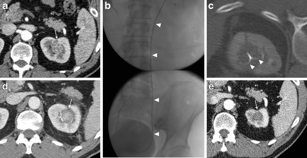
Teaching points: • Cryoablation, radiofrequency and microwave ablation have different advantages and disadvantages. • US, CT, fusion imaging, and CEUS increase an effective image-guidance. • Different patient positioning and external compression may increase procedure feasibility. • Hydrodissection and gas insufflation are useful to displace surrounding critical structures. • Cold pyeloperfusion can reduce the thermal damage to the collecting system.

Keywords: Image guidance; Image-guided tumour ablation; Protective manoeuvres; Renal ablation; Renal tumours.


Figures

Fig. 1
Fig. 1
Treatment of a right renal tumour with the application of US/CT fusion imaging. a Contrast enhanced CT demonstrating a right renal tumour (arrow). b needle insertion was performed with fusion of real-time US with the preacquired CT that allowed the precise positioning of the microwave antenna (arrowhead) at the level of the tumour (arrow). c CT acquisition immediately after antenna insertion confirmed the correct positioning of the device (arrow) in the desired position. d contrast enhanced CT 24 h after treatment demonstrated the correct ablation of the tumour (arrow)
Fig. 2
Fig. 2
Microwave ablation of a left posterior renal cell carcinoma close to the colon with hydrodissection. a Contrast enhanced CT demonstrating a left posterior renal cell carcinoma (white arrow) to be treated with percutaneous thermal ablation. b Contrast enhanced CT performed the day of the procedure. To achieve a safe path to the tumour the patient is placed in prone position with external compression. In this position, the colon (asterisks) appears to be close to the tumour to be treated (white arrow). c fusion of contrast enhanced CT and real-time US allowed for the identification of the tumour to be treated with US (white arrow = tumour; asterisks = colon). d under US guidance a small spinal needle (arrowheads) is placed in between the tumour (white arrow) and colon (asterisks) and fluid (hash) is injected. e A CT scan performed after fluid (hash) injection confirmed the correct displacement of the colon (asterisks) from the target tumour (white arrow) (arrowhead = spinal needle). f CT scan demonstrating the insertion of the microwave antenna (arrowhead) into the renal tumour (white arrow) (asterisks = colon; hash = injected fluid). g Contrast-enhanced CT performed the day after treatment demonstrating the complete ablation of the renal tumour (white arrow)without complications (asterisks = colon)
Fig. 3
Fig. 3
Treatment of an anterior left renal tumour in close proximity with a bowel loop. a CT performed the day of treatment showing the close proximity of the tumour to be treated (white arrow) with a bowel loop (asterisks). Note the external compression to obtain the desired position of the patient for an easy access to the target tumour. b multiplanar reconstruction of the CT images showing the close proximity of the tumour (white arrow) with a bowel loop (asterisks). c A CT scan performed after fluid (hash) injection confirmed the correct displacement of the colon (asterisks) from the target tumour (white arrow) (arrowhead = spinal needle). d CT scan demonstrating the insertion of the microwave antenna (arrowhead) into the renal tumour (white arrow). e Contrast-enhanced CT performed immediately after treatment demonstrating the complete ablation of the renal tumour (white arrow) without complications
Fig. 4
Fig. 4
Treatment of a right renal tumour with image guided thermal ablation with the assistance of CO2 injection. a contrast enhanced CT demonstrating an anterior right renal tumour close to the colon (arrow). b patient was positioned in lateral decubitus and a small needle was inserted in between the tumour and the colon (arrow). c CO2 was injected through the small needle (arrow) in order to achieve colon displacement. d an umbrella-shaped radiofrequency electrode (arrow) was inserted into the tumour from the opposite site of the colon, and slight traction made in order to displace the organ. e a contrast enhanced CT performed 24 h after the ablation demonstrated good ablation of the tumour (arrow) without complications
Fig. 5
Fig. 5
Treatment of a left kidney central tumour with renal sinus extension. a contrast enhanced CT demonstrating a centrally located tumour (white arrow) with extension in close proximity of the renal sinus. b retrograde pyeloperfusion was performed through a single j stent (arrowheads) placed endoscopically the day of the ablation. c CT scan after insertion on a radiofrequency electrode (arrowheads) into the tumour. d contrast enhanced CT performed immediately after the ablation demonstrated the complete ablation of the tumour (white arrowhead) without complications. e Contrast enhanced CT 2 years after the ablation demonstrating complete ablation with tumour shrinkage and no complications

References

    1. Hancock JB, Georgiades CS (2016) Kidney cancer. Cancer J. 22:387–392 - PubMed
    1. Mirzaei M, Pournamdar Z, Salehiniya H. Epidemiology and trends in incidence of kidney cancer in Iran. Asian Pacific J Cancer Prev. 2015;16:5859–5861. doi: 10.7314/APJCP.2015.16.14.5859. - DOI - PubMed
    1. Chow W, Dong L, Devesa S. Epidemiology and risk factors for kidney cancer. Nat Rev Urol. 2010;7:245–257. doi: 10.1038/nrurol.2010.46. - DOI - PMC - PubMed
    1. Herr H. A history of Partial nephrectomy for renal tumors. J Urol. 2005;173:705–708. doi: 10.1097/01.ju.0000146270.65101.1d. - DOI - PubMed
    1. Pierorazio PM, Johnson MH, Patel HD, et al. Management of Renal Masses and Localized Renal Cancer. Comp Eff Rev. 2016;167:1–20. - PubMed