. 2017 Sep;148(3):229-238.
doi: 10.1007/s00418-017-1578-0. Epub 2017 May 12.

Retinoic acid regulates cell-shape and -death of E-FABP (FABP5)-immunoreactive septoclasts in the growth plate cartilage of mice

Affiliations

Retinoic acid regulates cell-shape and -death of E-FABP (FABP5)-immunoreactive septoclasts in the growth plate cartilage of mice

Yasuhiko Bando et al. Histochem Cell Biol. 2017 Sep.

Abstract

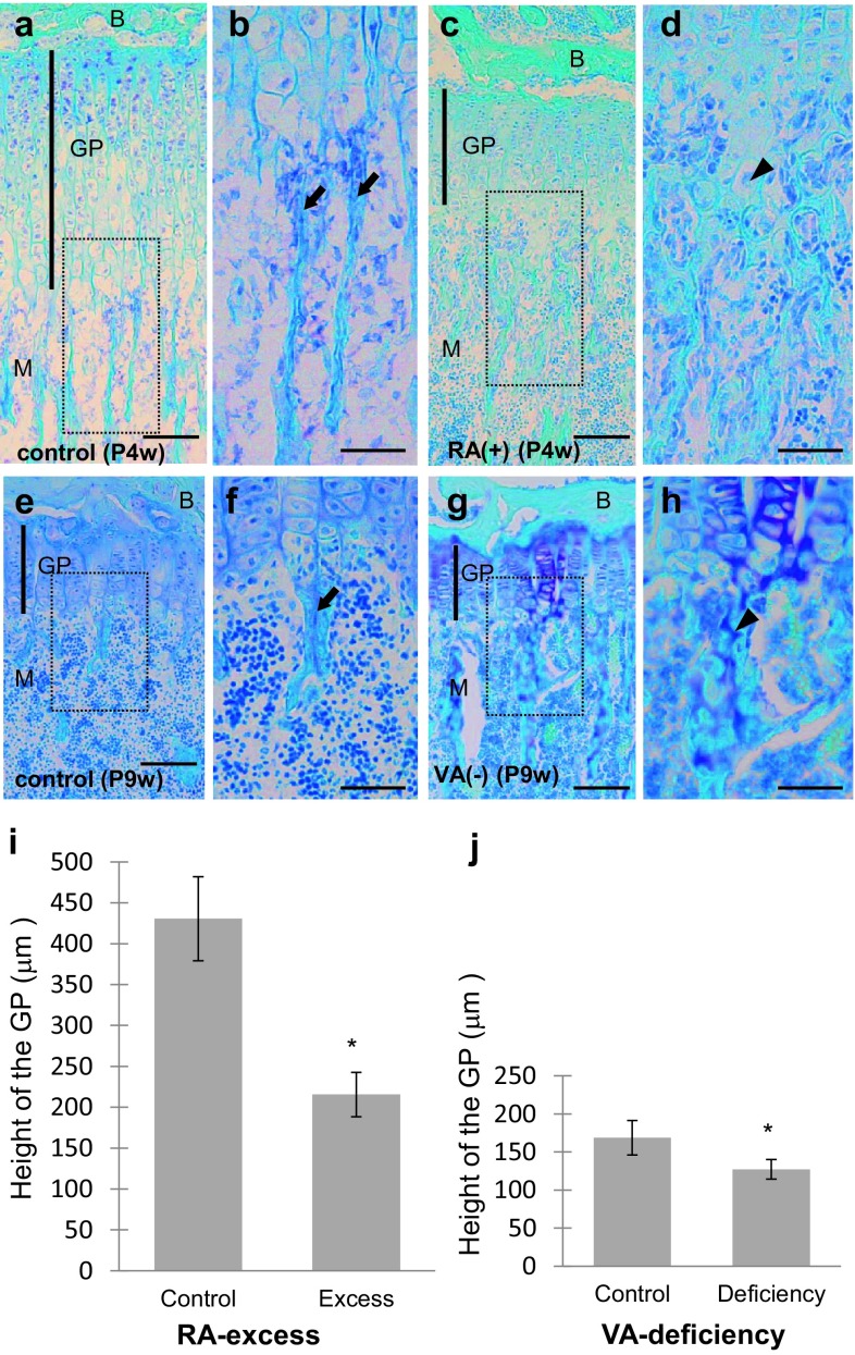
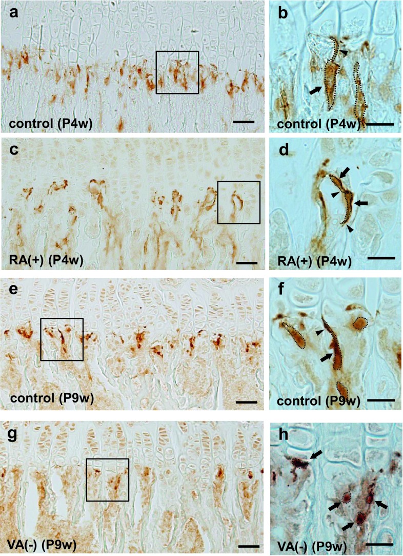
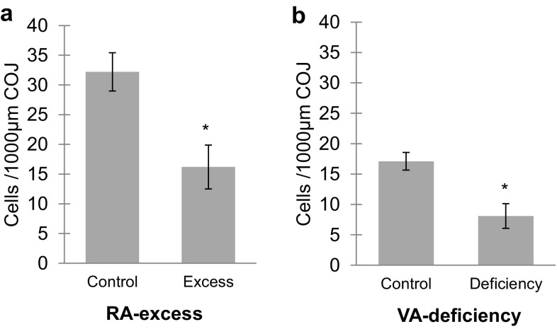
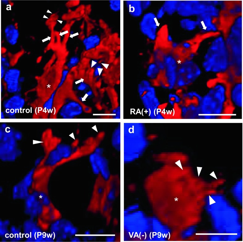
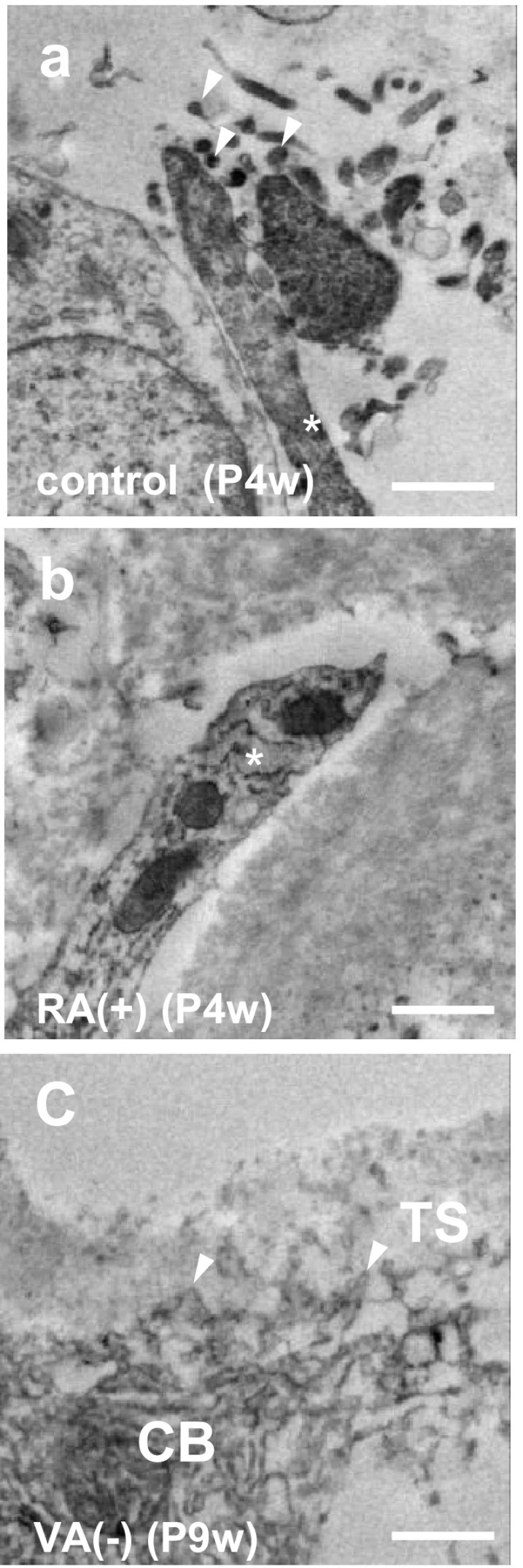
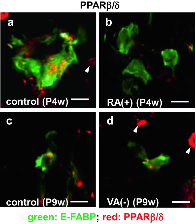
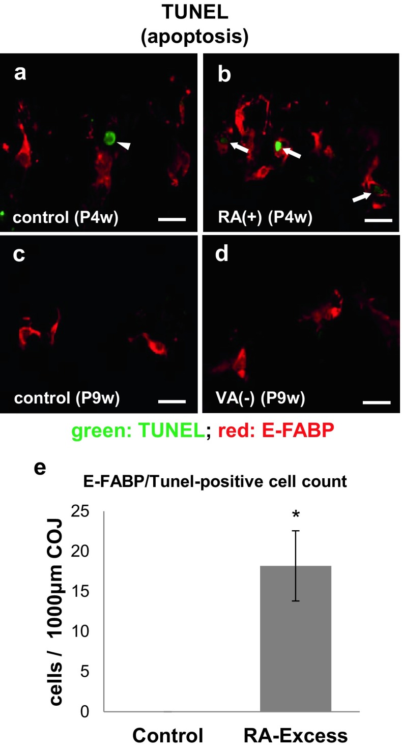
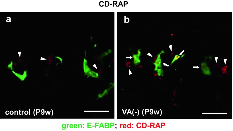
Septoclasts, which are mononuclear and spindle-shaped cells with many processes, have been considered to resorb the transverse septa of the growth plate (GP) cartilage at the chondro-osseous junction (COJ). We previously reported the expression of epidermal-type fatty acid-binding protein (E-FABP, FABP5) and localization of peroxisome proliferator-activated receptor (PPAR)β/δ, which mediates the cell survival or proliferation, in septoclasts. On the other hand, retinoic acid (RA) can bind to E-FABP and is stored abundantly in the GP cartilage. From these information, it is possible to hypothesize that RA in the GP is incorporated into septoclasts during the cartilage resorption and regulates the growth and/or death of septoclasts. To clarify the mechanism of the cartilage resorption induced by RA, we administered an overdose of RA or its precursor vitamin A (VA)-deficient diet to young mice. In mice of both RA excess and VA deficiency, septoclasts decreased in the number and cell size in association with shorter and lesser processes than those in normal mice, suggesting a substantial suppression of resorption by septoclasts in the GP cartilage. Lack of PPARβ/δ-expression, TUNEL reaction, RA receptor (RAR)β, and cellular retinoic acid-binding protein (CRABP)-II were induced in E-FABP-positive septoclasts under RA excess, suggesting the growth arrest/cell-death of septoclasts, whereas cartilage-derived retinoic acid-sensitive protein (CD-RAP) inducing the cell growth arrest or morphological changes was induced in septoclasts under VA deficiency. These results support and do not conflict with our hypothesis, suggesting that endogenous RA in the GP is possibly incorporated in septoclasts and utilized to regulate the activity of septoclasts resorbing the GP cartilage.

Keywords: E-FABP; Growth plate; Immunohistochemistry; Mouse; Retinoic acid; Septoclast.


Conflict of interest statement

Conflict of interest

The authors declare that they have no conflict of interest.

Statement on the welfare of animals

All animal procedures were in accordance with the Guidelines for care and use of Laboratory Animals of Meikai University School of Dentistry, and these experiments were approved by Meikai University Animal Ethics Committee (A1602).

Funding

This work was supported by JSPS KAKENHI Grant Numbers JP16K11457 to Y. B. and JP26462796 to O. A. and Meikai University Miyata Research Grant (2016 A to Y. B.).

Figures

Fig. 1
Fig. 1
The growth plate (GP) in mouse tibiae. Light micrographs of toluidine blue-stained sections at lower (a, c, e, g) and higher (b, d, f, h squares in a, c, e, and g, respectively) magnifications and graphs (i, j) showing the effects of retinoic acid (RA)-excessive administration for 4-week-old (P4w) (c, d; a, b: control) or vitamin A (VA)-deficient diet (g, h, 6 weeks from P3w; e, f: control) on the GP of the proximal tibiae of mice. B epiphyseal bone, M metaphysis, arrows trabeculae, arrowheads cartilage, scale bars 100 μm; mean ± SD; *P < 0.01 (n = 50)
Fig. 2
Fig. 2
Chodro-osseous junction (COJ) of the growth plate in mouse tibiae. Light micrographs at lower (a, c, e, g) and higher (b, d, f, h squares in a, c, e, and g, respectively) magnifications at the COJ of the GP in mouse proximal tibiae of P4w control (a, b), P4w RA excess (c, d), P9w control (e, f), and P9w VA deficiency (g, h). Dotted line outline of each E-FABP-immunopositive septoclast; arrows septoclastic cell bodies; arrowheads septoclastic processes; scale bars 50 μm (a, c, e, g) or 20 μm (b, d, f, h)
Fig. 3
Fig. 3
Graph showing numerical changes of E-FABP-immunoreactive septoclasts. The cells were counted at the COJ in an area of 1000-μm width ×200-μm height in the GP of mouse proximal tibiae of RA excess (a) or VA deficiency (b). Values are shown as mean ± SD; *P < 0.01 (n = 10)
Fig. 4
Fig. 4
Three-dimensional (3D) images of E-FABP-immunoreactive septoclasts. Confocal laser-scanning microscopy of the 3D reconstruction of E-FABP-immunoreactive (red) septoclasts (ad) at the COJ of the GP in mouse proximal tibiae of P4w control (a), P4w RA excess (b), P9w control (c), and P9w VA deficiency (d). Nuclei are stained with DAPI (bluish purple). Asterisks septoclastic bodies, arrows septoclastic processes, arrowheads microvilli (a) or short cytoplasmic spines (d); scale bars 10 μm
Fig. 5
Fig. 5
Process apex of septoclasts. Immunoelectron micrographs of E-FABP-immunoreactive septoclasts at the COJ of the GP of mouse proximal tibiae of P4w control (a), P4w RA excess (b), and P9w VA deficiency (c). Asterisks septoclastic processes; arrowheads: microvilli (a) or short spines of septoclastic cell body (c); CB septoclastic cell body, TS transverse septum; scale bars 1 μm
Fig. 6
Fig. 6
Double staining for E-FABP plus PPARβ/δ in septoclasts. Confocal laser-scanning micrographs of double immunostaining for E-FABP (green) plus PPARβ/δ (red) at the COJ of the GP in mouse proximal tibiae of P4w control (a), P4w RA excess (b), P9w control (c), and P9w VA deficiency (d). Arrowheads PPARβ/δ-immunoreactive chondrocytes; scale bars 10 μm
Fig. 7
Fig. 7
E-FABP-immunoreactivity and apoptosis in septoclasts. Confocal laser-scanning micrographs of double staining for E-FABP (red) plus TUNEL reaction (green) at the COJ of the GP in mouse proximal tibiae of P4w control (a), P4w RA excess (b), P9w control (c), and P9w VA deficiency (d) and the graph (e) showing the significant increase of simultaneously E-FABP-immunoreactive and TUNEL-positive apoptotic cells at the COJ of the GP in mouse proximal tibiae of RA excess in an area of 1000-μm width ×200-μm height as compared to the control. Arrows: TUNEL-positive septoclasts; arrow head: TUNEL-positive chondrocyte; scale bars 20 μm; mean ± SD; *P < 0.01 (n = 10)
Fig. 8
Fig. 8
Double staining for E-FABP plus RARβ (a, b) and for CRABP-II plus cathepsin B (c, d). Confocal laser-scanning micrographs of double staining for E-FABP (green, a, b) plus RARβ (red, a, b), and for CRABP-II (green c, d) plus cathepsin B (red c, d) at the COJ of the GP in mouse proximal tibiae of P4w control (a, c) and P4w RA excess (b, d). Arrows RAR-immunoreactive septoclasts; arrowheads CRABP-II-immunoreactive septoclasts; scale bars 20 μm
Fig. 9
Fig. 9
Double staining for E-FABP plus CD-RAP in septoclasts. Confocal laser-scanning micrographs of double staining for E-FABP (green) plus CD-RAP (red) at the COJ of the GP in mouse proximal tibiae of P9w control (a) and P9w VA deficiency (b). Arrows CD-RAP-immunoreactive septoclasts; arrowheads CD-RAP-immunoreactivity in chondrocytes; scale bars 20 μm

References

    1. Al Tanoury Z, Piskunov A, Rochette-Egly C. Vitamin A and retinoid signaling: genomic and nongenomic effects. J Lipid Res. 2013;54:1761–1775. doi: 10.1194/jlr.R030833. - DOI - PMC - PubMed
    1. Amano O, Kudo Y, Shimada M, Wakayama T, Yamamoto M, Iseki S. Transient occurrence of 27 kDa heat-shock protein in the terminal tubule cells during postnatal development of the rat submandibular gland. Anat Rec. 2001;264:358–366. doi: 10.1002/ar.10023. - DOI - PubMed
    1. Bando Y, Yamamoto M, Sakiyama K, Inoue K, Takizawa S, Owada Y, Iseki S, Kondo H, Amano O. Expression of epidermal fatty acid binding protein (E-FABP) in septoclasts in the growth plate cartilage of mice. J Mol Histol. 2014;45:507–518. doi: 10.1007/s10735-014-9576-1. - DOI - PubMed
    1. Blesch A, Bosserhoff AK, Apfel R, Behl C, Hessdoerfer B, Schmitt A, Jachimczak P, Lottspeich F, Buettner R, Bogdahn U. Cloning of a novel malignant melanoma-derived growth-regulatory protein, MIA. Cancer Res. 1994;54:5695–5701. - PubMed
    1. De Luca F, Uyeda JA, Mericq V, Mancilla EE, Yanovski JA, Barnes KM, Zile MH, Baron J. Retinoic acid is a potent regulator of growth plate chondrogenesis. Endocrinology. 2000;141:346–353. doi: 10.1210/endo.141.1.7283. - DOI - PubMed

MeSH terms

LinkOut - more resources