Review
. 2017:84:103-137.
doi: 10.1007/978-3-319-53047-5_4.

Cytoskeletal Proteins in Caulobacter crescentus: Spatial Orchestrators of Cell Cycle Progression, Development, and Cell Shape

Affiliations
Review

Cytoskeletal Proteins in Caulobacter crescentus: Spatial Orchestrators of Cell Cycle Progression, Development, and Cell Shape

Kousik Sundararajan et al. Subcell Biochem. 2017.

Abstract

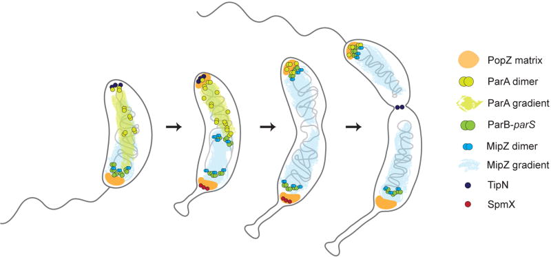
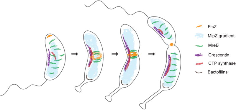
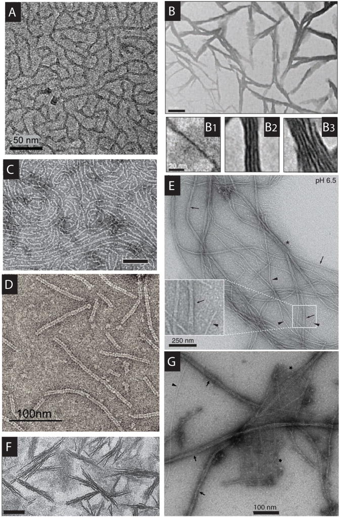
Caulobacter crescentus, an aquatic Gram-negative α-proteobacterium, is dimorphic, as a result of asymmetric cell divisions that give rise to a free-swimming swarmer daughter cell and a stationary stalked daughter. Cell polarity of vibrioid C. crescentus cells is marked by the presence of a stalk at one end in the stationary form and a polar flagellum in the motile form. Progression through the cell cycle and execution of the associated morphogenetic events are tightly controlled through regulation of the abundance and activity of key proteins. In synergy with the regulation of protein abundance or activity, cytoskeletal elements are key contributors to cell cycle progression through spatial regulation of developmental processes. These include: polarity establishment and maintenance, DNA segregation, cytokinesis, and cell elongation. Cytoskeletal proteins in C. crescentus are additionally required to maintain its rod shape, curvature, and pole morphology. In this chapter, we explore the mechanisms through which cytoskeletal proteins in C. crescentus orchestrate developmental processes by acting as scaffolds for protein recruitment, generating force, and/or restricting or directing the motion of molecular machines. We discuss each cytoskeletal element in turn, beginning with those important for organization of molecules at the cell poles and chromosome segregation, then cytokinesis, and finally cell shape.


Figures

Figure 1
Figure 1. Cytoskeletal elements involved in cell cycle progression
Swarmer cells (left, with polar flagellum) and newly divided stalked cells have a unipolar PopZ matrix and a single ParB focus (bound to parS) close to the old pole. ParA and MipZ bind DNA (gray line) non-specifically and localize as diffuse clouds enriched at the pole(s). In newborn cells, MipZ is concentrated at the old pole by ParB bound to PopZ whereas ParA is concentrated at the new pole by TipN. Following initiation of chromosome replication, a new PopZ matrix is assembled at the new pole. At the same time, PopZ at the old pole recruits SpmX to drive stalk biogenesis. As chromosome replication progresses, one of the ParB foci moves up the ParA concentration gradient towards the new pole, leading to an ori-ter-ori orientation of the chromosome. MipZ is segregated to the new pole with ParB-parS, leading to a MipZ concentration gradient with the minimum at approximately midcell. See text for more details.
Figure 2
Figure 2. Cytoskeletal regulation of cell shape and morphogenesis
In swarmer cells, dynamic clusters of MreB are distributed along the length of the cell and direct cell elongation. FtsZ is localized to the new cell pole in swarmers, but due to the dynamic relocation of the MipZ concentration maxima to the poles upon centromere segregation, FtsZ is displaced from the new pole and reassembled at midcell to form the Z-ring. Following Z-ring assembly, MreB is recruited to the midcell where it drives midcell elongation along with FtsZ. Finally, following the arrival of late division proteins, MreB is displaced from the midcell into dispersed clusters. Crescentin and CTP synthase work synergistically to direct cell curvature. Crescentin forms short structures at the inner cell curvature in swarmer cells, which elongate as the cell grows until these crescentin filaments reach the cell poles. CTP synthase forms shorter, predominantly cytoplasmic filaments in swarmer cells and longer, membrane-associated structures in stalked cells. Bactofilins assemble into a scaffold in early stalked cells at the base of the stalk and regulate pole morphogenesis.
Figure 3
Figure 3. Function and localization of C. crescentus cytoskeleton
(A) Phase contrast image of wild-type C. crescentus cells (Unpublished results). Figures (B) – (H) represent morphological changes upon deletion, depletion or overexpression of individual cytoskeletal elements. Scale bars = 2 µm. (B) DIC image of ΔpopZ cells. Arrows highlight minicell formation (Ebersbach et al. 2008). (C) Phase contrast image of cells overexpressing parA (Mohl & Gober 1997). (D) Phase contrast image of cells depleted of FtsZ for 4.5 hours (Sundararajan et al. 2015). (E) Phase contrast image of cells depleted of MreB for 10 hours (Figge et al. 2004). (F) DIC image of ΔcreS cells (Ausmees et al. 2003). (G) Phase contrast image of cells depleted of CTP synthase (Ingerson-Mahar et al. 2010). (H) Phase contrast image of cells overexpressing bacA-venus (Kühn et al. 2009). Figures (I) – (O) represent localization pattern of individual cytoskeletal proteins. (I) Epifluorescence microscopy showing polar localization of PopZ-YFP (in red overlaid on DIC images) in synchronized cells; I1 – early swarmer with unipolar PopZ localization, I2 – Pre-divisional cell with bipolar PopZ localization, I3 – Bipolar PopZ localization in cell undergoing cytokinesis. Scale bar = 2µm (Ebersbach et al. 2008). (J) PALM image of ParA-Dendra2 expressed from native locus (J1) or under the control of Pxyl promoter (J2). Scale bar = 1 µm (Lim et al. 2014). (K) HT-PALM image of FtsZ-Dendra2 in a predivisional cell; (K1) and (K2) represent longitudinal and cross-sectional views of the Z-ring respectively. Scale bar = 500 nm (Holden et al. 2014). (L) PALM image of eYFP-MreB. Scale bar = 300 nm (Biteen et al. 2008). (M) Immunofluorescence microscopy image showing CreS-FLAG (red) and the nucleoid stained with DAPI (blue). Scale bar = 2 µm (Ausmees et al. 2003). (N) Epifluorescence microscopy of cells expressing mChy-CtpS (red overlaid on phase contrast image). Scale bar = 2 µm (Ingerson-Mahar et al. 2010). (O) Merged image showing localization of BacA-eCFP and BacB-Venus. Scale bar = 2 µm (Kühn et al. 2009).
Figure 4
Figure 4. Diverse cytoskeletal structures observed in vivo
A subset of the cytoskeletal elements described in C. crescentus has been imaged at high resolution in vivo using electron cryotomography (ECT). ECT images of C. crescentus cells showing (A) Ribosome-free zone associated with the PopZ matrix at the old cell pole (highlighted by dotted line) (Bowman et al. 2010), (B) FtsZ protofilaments (red) assembled into a ring proximal to the inner membrane (blue) at the site of division (Z. Li et al. 2007), (C) Filaments formed on overproduction of crescentinΔN27 (scale bar = 50 nm) (Cabeen et al. 2010), (D) CTP synthase ribbons assembled at the inner curvature of the cell proximal to the inner membrane (scale bar = 200 nm) (Briegel et al. 2006; Ingerson-Mahar et al. 2010) (E) Bactofilin scaffold assembled at the base of the stalk (scale bar = 50 nm) (Kühn et al. 2009).
Figure 5
Figure 5. Filaments and higher order structures formed by cytoskeletal proteins in vitro
Transmission electron microscopy (TEM) images of purified proteins reported to have been assembled into higher order structures in vitro. (A) PopZ filaments, scale bar = 50 nm (Bowman et al. 2008). (B) ParA filaments assembled in the presence of ATP, scale bar = 100 nm. (B1), (B2), (B3) show enlarged images of filament bundles, scale bar = 20 nm (Ptacin et al. 2010). (C) FtsZ protofilaments assembled in the presence of GTP, scale bar = 100 nm (Unpublished results). (D) Double protofilaments of MreB assembled on a lipid monolayer in the presence of ATP, scale bar = 100 nm (van den Ent et al. 2014). (E) Crescentin filaments (arrowheads) and bundles (arrows and asterisks) assembled at pH 6.5 in the absence of Mg2+, scale bar = 250 nm (Cabeen et al. 2010). (F) Filaments formed by E. coli CTP synthase, scale bar = 100 nm (Ingerson-Mahar et al. 2010). (G) Filaments (arrows) and 2D-crystalline sheets (asterisks) formed by BacA, scale bar = 100 nm (Vasa et al. 2015). Note that the identities of structures seen in some TEM images published in papers on cytoskeletal filaments have been questioned (Ghosal et al, 2014, supplementary file; Griffith & Bonner, 1973), since contaminating cellulose fibers and thin uranyl acetate crystals may each be mistaken for protein polymers; here, the FtsZ and MreB filaments in C and D appear white, as expected for protein embedded in negative stain, and are also unmistakably identifiable from longitudinal repeats corresponding to the sizes of the monomers, but the EM evidence for filament formation by other proteins is weaker.

Similar articles

Cited by

References

    1. Aaron M, et al. The tubulin homologue FtsZ contributes to cell elongation by guiding cell wall precursor synthesis in Caulobacter crescentus. Molecular Microbiology. 2007;64(4):938–952. - PubMed
    1. Ausmees N, Kuhn JR, Jacobs-Wagner C. The bacterial cytoskeleton: an intermediate filament-like function in cell shape. Cell. 2003;115(6):705–713. - PubMed
    1. Banigan EJ, et al. Filament depolymerization can explain chromosome pulling during bacterial mitosis. PLoS computational biology. 2011;7(9):e1002145. - PMC - PubMed
    1. Barry RM, et al. Large-scale filament formation inhibits the activity of CTP synthetase. eLife. 2014;3:e03638. - PMC - PubMed
    1. Bartosik AA, et al. ParB deficiency in Pseudomonas aeruginosa destabilizes the partner protein ParA and affects a variety of physiological parameters. Microbiology (Reading, England) 2009;155(Pt 4):1080–1092. - PMC - PubMed

MeSH terms

LinkOut - more resources