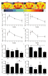
Identification of changes in neuronal function as a consequence of aging and tauopathic neurodegeneration using a novel and sensitive magnetic resonance imaging approach
- PMID: 28500878
- PMCID: PMC5524451
- DOI: 10.1016/j.neurobiolaging.2017.04.007
Identification of changes in neuronal function as a consequence of aging and tauopathic neurodegeneration using a novel and sensitive magnetic resonance imaging approach
Abstract
Tauopathies, the most common of which is Alzheimer's disease (AD), constitute the most crippling neurodegenerative threat to our aging population. Tauopathic patients have significant cognitive decline accompanied by irreversible and severe brain atrophy, and it is thought that neuronal dysfunction begins years before diagnosis. Our current understanding of tauopathies has yielded promising therapeutic interventions but have all failed in clinical trials. This is partly due to the inability to identify and intervene in an effective therapeutic window early in the disease process. A major challenge that contributes to the definition of an early therapeutic window is limited technologies. To address these challenges, we modified and adapted a manganese-enhanced magnetic resonance imaging (MEMRI) approach to provide sensitive and quantitative power to detect changes in broad neuronal function in aging mice. Considering that tau tangle burden correlates well with cognitive impairment in Alzheimer's patients, we performed our MEMRI approach in a time course of aging mice and an accelerated mouse model of tauopathy. We measured significant changes in broad neuronal function as a consequence of age, and in transgenic mice, before the deposition of bona fide tangles. This MEMRI approach represents the first diagnostic measure of neuronal dysfunction in mice. Successful translation of this technology in the clinic could serve as a sensitive diagnostic tool for the definition of effective therapeutic windows.
Keywords: Alzheimer; MEMRI; Manganese; Tangles; Tau; rTg4510.
Copyright © 2017 Elsevier Inc. All rights reserved.
Conflict of interest statement
All authors consent publication of this manuscript. There are no competing interests. Funding for this work came from the University of Kentucky Alzheimer’s Disease Center (UK-ADC), which is supported by NIH/NIA P30 AG028383. JFA, SNF, AI, RAC. SEM, EM, DL, GKN, and EM, were supported by NIH/NINDS 1R01 NS091329-01, Alzheimer’s Association NIRG-14-322441, NIH/NCATS 5UL1TR000117-04, NIH/NIGMS 5P30GM110787, Department of Defense AZ140097, the University of Kentucky Epilepsy Center (EpiC), NIH/NIA P30 AG028383, and NIH/NIMHD L32 MD009205-01. We acknowledge Dr. Donna Wilcock for use of the AxioScan, Dr. Peter Davies for the MC1 antibody, and Dr. Frederick Schmitt for comments on this manuscript and insights into these experiments. Author contributions are as follows: JFA designed the study. SNF, AI, SEM, EM, and DL performed MRI. MV developed analysis program for MEMRI and MV, DL, SEM, and RAC analyzed MEMRI data. AI and EM performed mouse husbandry and with SNF, tissue collection. SNF performed all IHC and analysis. SNF, MV and JFA interpreted data and wrote the paper. Data and materials will be made widely available upon reasonable request to the corresponding author.
Figures


Similar articles
-
Selective Disruption of Inhibitory Synapses Leading to Neuronal Hyperexcitability at an Early Stage of Tau Pathogenesis in a Mouse Model.J Neurosci. 2020 Apr 22;40(17):3491-3501. doi: 10.1523/JNEUROSCI.2880-19.2020. Epub 2020 Apr 7. J Neurosci. 2020. PMID: 32265258 Free PMC article.
-
Broad Kinase Inhibition Mitigates Early Neuronal Dysfunction in Tauopathy.Int J Mol Sci. 2021 Jan 26;22(3):1186. doi: 10.3390/ijms22031186. Int J Mol Sci. 2021. PMID: 33530349 Free PMC article.
-
Non-invasive, in vivo monitoring of neuronal transport impairment in a mouse model of tauopathy using MEMRI.Neuroimage. 2013 Jan 1;64:693-702. doi: 10.1016/j.neuroimage.2012.08.065. Epub 2012 Aug 31. Neuroimage. 2013. PMID: 22960250 Free PMC article.
-
[Development of imaging-based diagnostic procedures for brain protein aging using a mouse model of tauopathy].Nihon Yakurigaku Zasshi. 2018;152(1):4-9. doi: 10.1254/fpj.152.4. Nihon Yakurigaku Zasshi. 2018. PMID: 29998952 Review. Japanese.
-
Tau imaging in the study of ageing, Alzheimer's disease, and other neurodegenerative conditions.Curr Opin Neurobiol. 2016 Feb;36:43-51. doi: 10.1016/j.conb.2015.09.002. Epub 2015 Sep 20. Curr Opin Neurobiol. 2016. PMID: 26397020 Review.
Cited by
-
Manganese-Enhanced Magnetic Resonance Imaging: Application in Central Nervous System Diseases.Front Neurol. 2020 Feb 25;11:143. doi: 10.3389/fneur.2020.00143. eCollection 2020. Front Neurol. 2020. PMID: 32161572 Free PMC article. Review.
-
In vivo tracing of the ascending vagal projections to the brain with manganese enhanced magnetic resonance imaging.Front Neurosci. 2023 Sep 15;17:1254097. doi: 10.3389/fnins.2023.1254097. eCollection 2023. Front Neurosci. 2023. PMID: 37781260 Free PMC article.
-
Activity-induced MEMRI cannot detect functional brain anomalies in the APPxPS1-Ki mouse model of Alzheimer's disease.Sci Rep. 2019 Feb 4;9(1):1140. doi: 10.1038/s41598-018-37980-y. Sci Rep. 2019. PMID: 30718666 Free PMC article.
-
A new opportunity for MEMRI.Aging (Albany NY). 2017 Aug 28;9(8):1855-1856. doi: 10.18632/aging.101283. Aging (Albany NY). 2017. PMID: 28854148 Free PMC article. No abstract available.
-
Neuroimmune Tau Mechanisms: Their Role in the Progression of Neuronal Degeneration.Int J Mol Sci. 2018 Mar 23;19(4):956. doi: 10.3390/ijms19040956. Int J Mol Sci. 2018. PMID: 29570615 Free PMC article. Review.
References
-
- Abisambra J, Jinwal UK, Miyata Y, Rogers J, Blair L, Li X, Seguin SP, Wang L, Jin Y, Bacon J, Brady S, Cockman M, Guidi C, Zhang J, Koren J, Young ZT, Atkins CA, Zhang B, Lawson LY, Weeber EJ, Brodsky JL, Gestwicki JE, Dickey CA. Allosteric heat shock protein 70 inhibitors rapidly rescue synaptic plasticity deficits by reducing aberrant tau. Biological psychiatry. 2013;74(5):367–374. - PMC - PubMed
-
- Abisambra JF, Blair LJ, Hill SE, Jones JR, Kraft C, Rogers J, Koren J, 3rd, Jinwal UK, Lawson L, Johnson AG, Wilcock D, O’Leary JC, Jansen-West K, Muschol M, Golde TE, Weeber EJ, Banko J, Dickey CA. Phosphorylation dynamics regulate Hsp27-mediated rescue of neuronal plasticity deficits in tau transgenic mice. The Journal of neuroscience: the official journal of the Society for Neuroscience. 2010a;30(46):15374–15382. - PMC - PubMed
-
- Alzheimer’s A. 2016 Alzheimer’s disease facts and figures. Alzheimer’s & dementia: the journal of the Alzheimer’s Association. 2016;12(4):459–509. - PubMed
-
- Antkowiak PF, Vandsburger MH, Epstein FH. Quantitative pancreatic beta cell MRI using manganese-enhanced Look-Locker imaging and two-site water exchange analysis. Magnetic resonance in medicine: official journal of the Society of Magnetic Resonance in Medicine/Society of Magnetic Resonance in Medicine. 2012;67(6):1730–1739. - PubMed
Publication types
MeSH terms
Substances
Grants and funding
LinkOut - more resources
Full Text Sources
Other Literature Sources
Medical
Molecular Biology Databases

