. 2017 May 1:211:251-260.
doi: 10.1016/j.jss.2016.12.028. Epub 2016 Dec 29.

Direct orthotopic implantation of hepatic organoids

Affiliations

Direct orthotopic implantation of hepatic organoids

Vivian X Zhou et al. J Surg Res. .

Abstract

Background: Liver organoids show potential for development as a tissue replacement therapy for patients with end-stage liver disease, but efficient methods for introducing organoids into host livers have not been established. In this study, we aimed to develop a surgical technique to implant hepatic organoids into the liver and assess their engraftment.

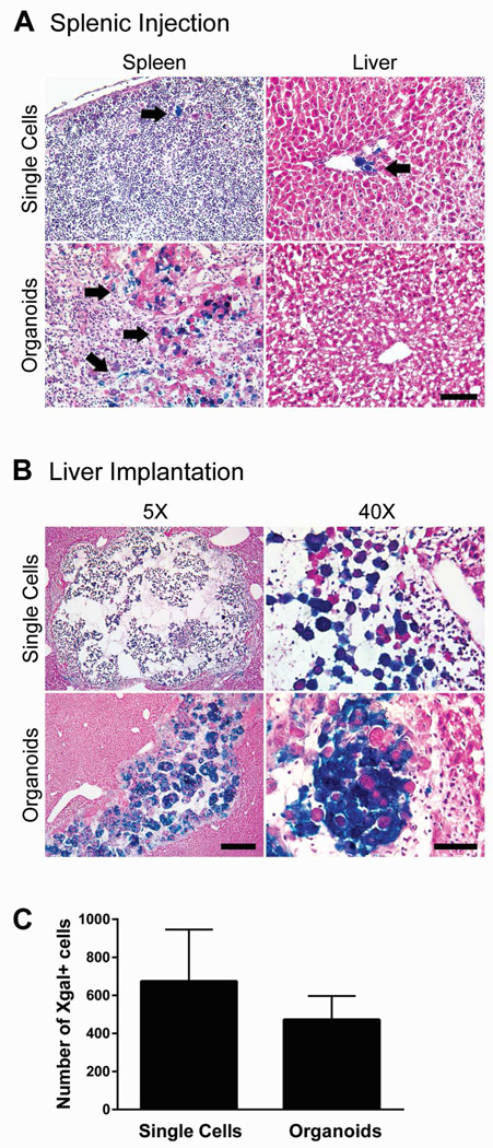
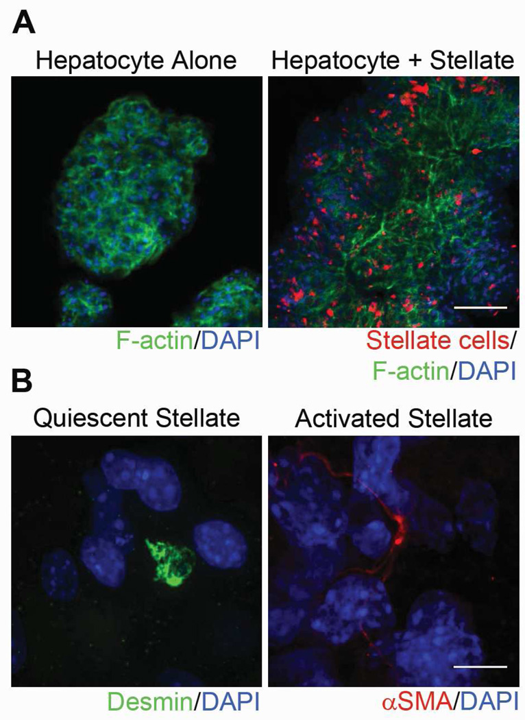
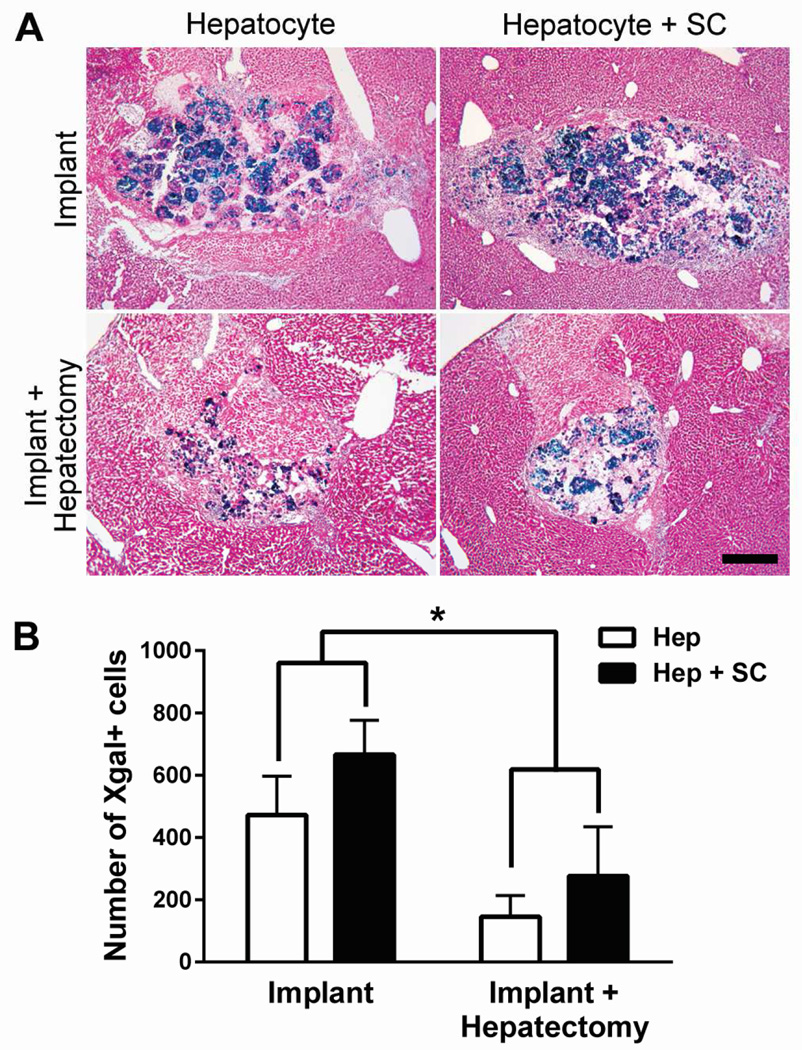
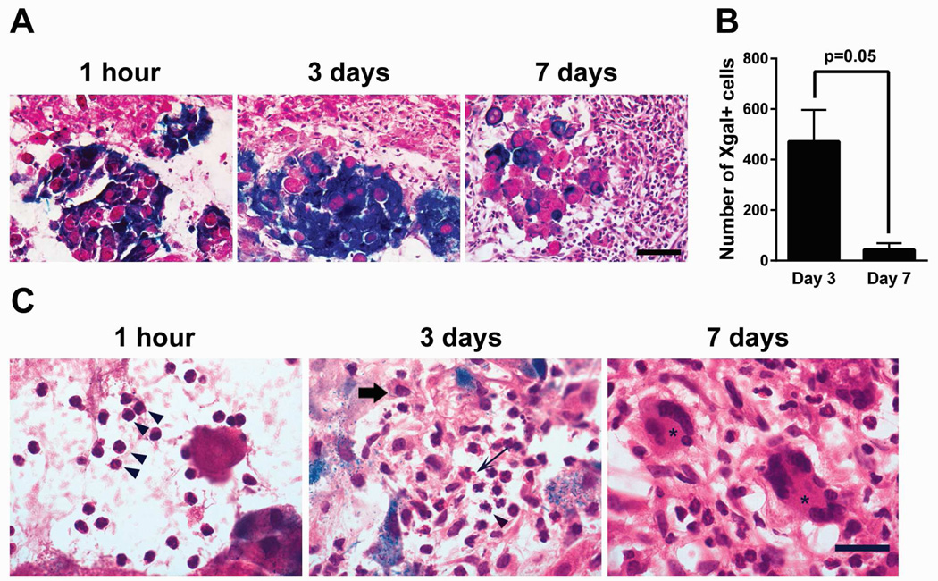
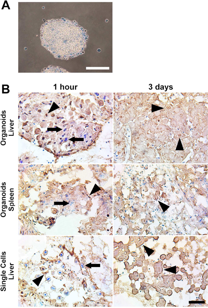
Methods: Donor hepatocytes were isolated from ROSA26 C57BL/6 mice, so that engrafted cells, when implanted into wild-type mice, could be easily identified by X-gal staining. Hepatic organoids were generated by three-dimensional culture in rotating wall vessel bioreactors. We qualitatively and quantitatively compared organoid engraftment to that of single-cell hepatocyte transplants. In addition, we determined the effect of adding stellate cells to hepatocytes to form co-aggregated organoids and the effect of partial hepatectomy of the host liver on organoid engraftment.

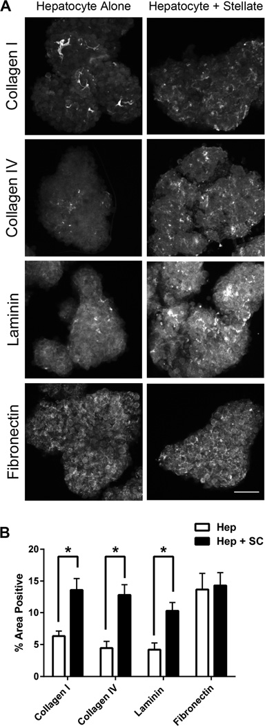
Results: Direct orthotopic implantation of hepatic organoids within a hepatotomy site resulted in local engraftment of exogenous hepatocytes with limited durability. Hepatocyte-stellate cell organoids produced more extracellular matrix but did not significantly improve engraftment compared with hepatocyte-alone organoids. Partial hepatectomy of the host liver led to significantly decreased engraftment of organoids. Survival of organoids was limited by the presence of apoptotic hepatocytes within organoids as early as 1 h after implantation. Organoids eventually became necrotic and elicited a chronic inflammatory giant cell reaction similar to a foreign body response.

Conclusions: With additional organoid and host factor optimization, direct orthotopic implantation of hepatic organoids may be an approach to introduce large numbers of exogenous hepatocytes into recipient livers.

Keywords: Liver organoids; Three-dimensional culture; Tissue engineering; Transplantation.


Conflict of interest statement

Disclosure

The authors have no conflicts to disclose.

Figures

Figure 1
Figure 1
Liver organoids injected through the spleen remain trapped within the spleen, whereas liver organoids orthotopically implanted directly into the liver parenchyma engraft locally within the implanted liver lobe. (A) Representative images of spleen and liver from recipient mice transplanted with Xgal+ single-cell hepatocytes or organoids by injection through the spleen. Splenic injection of single cells showed rare retention of Xgal+ hepatocytes in the spleen and low efficiency engraftment near portal veins within the liver parenchyma (arrows). Transplanted organoids remained trapped within the spleen (arrows). Magnification 5x. Scale bar represents 400µm. (B) Representative images of livers that were directly implanted with single-cell hepatocytes or organoids. Implantation of either single cells or organoids showed local engraftment in the implanted liver lobe. Single cells were loosely dispersed within the hepatotomy site. Organoids retained their compact spheroid architecture. Scale bars represent 400µm (5x images) and 60µm (40x images). (C) Quantification of implanted hepatocytes by enumerating Xgal+ cells in single cell (n=9) versus organoid (n=6) orthotopic implantation. Error bars represent SEM.
Figure 2
Figure 2
Hepatocyte-stellate cell organoids demonstrate widespread distribution of stellate cells throughout the organoid and include both quiescent and activated stellate cells. (A) Representative confocal images of hepatocyte-alone and hepatocyte-stellate cell organoids. Organoids were stained with phalloidin to indicate F-actin (green) and DAPI to indicate nuclei (blue). Stellate cells were pretreated and identified with CellTracker CMTPX Red dye (red). Magnification 10x. Scale bar represents 100µm. (B) Representative confocal images of individual stellate cells within hepatocyte-stellate organoids show the presence of both quiescent and activated states. Quiescent stellate cells were identified via desmin (green) and round cell shape, whereas activated stellate cells were marked by αSMA (red) and elongated cell shape. Nuclei were stained with DAPI (blue). Magnification 100x. Scale bar represents 10µm.
Figure 3
Figure 3
Hepatocyte-stellate cell organoids show significant increases in several extracellular matrix (ECM) proteins compared to hepatocyte-alone organoids. (A) Representative confocal images of hepatocyte-stellate cell organoids demonstrated increased presence of collagen I, collagen IV, and laminin compared to hepatocyte-alone organoids, whereas levels of fibronectin were similar between the two organoid types. Magnification 10x. Scale bar represents 100µm. (B) Quantification of ECM components in hepatocyte-only (Hep) and hepatocyte-stellate (Hep + SC) organoids. Percent area positive for each ECM protein was determined by digital imaging analysis in at least 10 individual organoids per ECM component per group. *p<0.05 by two-tailed Student’s t-test. Error bars represent SEM.
Figure 4
Figure 4
Hepatocyte-stellate cell organoids show engraftment comparable to that of hepatocyte-alone organoids, whereas partial hepatectomy significantly reduces engraftment of both organoid types. (A) Representative images of liver sections demonstrating implantation outcomes of hepatocyte-alone (Hep) organoids with (n=9) and without (n=6) partial hepatectomy, in comparison with hepatocyte-stellate cell (Hep + SC) organoids with (n=3) and without (n=3) partial hepatectomy. Images are representative of at least 6 sections evaluated per mouse. Magnification 5x. Scale bar represents 400µm. (B) Quantification of implanted hepatocytes by enumerating Xgal+ cells through digital image analysis. *p<0.01 by two-way ANOVA. Two-way ANOVA analysis showed no significant interaction (p = 0.79) between the type of surgery (with or without hepatectomy) and the type of organoid (with or without stellate cells). The p-values for the type of surgery and the type of organoid are 0.008 and 0.19, respectively. Error bars represent SEM.
Figure 5
Figure 5
Inflammatory cells surrounding implanted organoids accumulate over time, starting with a predominantly neutrophilic infiltration and evolving into a chronic inflammatory reaction with multinucleated giant cells. (A) Representative images of host inflammatory response and hepatocyte organoids in liver sections at 1 hour (n=3), 3 days (n=6), and 7 days (n=7) after organoid implantation. Images are representative of at least 4 sections evaluated per mouse. Magnification 40x. Scale bar represents 60µm. (B) Time course of Xgal+ cells detected at organoid implantation sites comparing 3 days (n=6) and 7 days (n=7) after implantation. p=0.05 by two-tailed Student’s t-test. Error bars represent SEM. (C) High-powered morphological analysis of the inflammatory infiltrates demonstrated the presence of predominantly neutrophils (arrowheads) at 1 hour after implantation. By 3 days, the expanded inflammatory infiltrate included recruitment of neutrophils (arrowhead), eosinophils (thin arrow), and macrophages (thick arrow). By 7 days, a chronic inflammatory reaction had developed with numerous multinucleated giant cells (asterisk). Magnification 60x. Scale bar represents 20µm.
Figure 6
Figure 6
Hepatocytes within viable organoids implanted into the liver parenchyma show early apopotosis and become necrotic by 3 days. (A) Hepatocyte organoids prior to implantation are viable and exclude trypan blue. Hepatocytes that remain as single cells in 3D culture are dead and trypan blue positive. Scale bar represents 200µm. (B) Hepatocyte organoids or freshly isolated single-cell hepatocytes were implanted into the liver or spleen. At 1 hour and 3 days post-implantation, TUNEL staining was performed to detect apoptotic nuclei (brown nuclei, indicated by arrowheads), and counterstained with methyl green for viable nuclei (blue nuclei, indicated by arrows). By 3 days after implantation, most hepatocytes were anucleated with washed-out cytoplasm, consistent morphologically with necrosis. Scale bar represents 100µm.

References

    1. Lancaster MA, Knoblich JA. Organogenesis in a dish: modeling development and disease using organoid technologies. Science. 2014;345:1247125. - PubMed
    1. 12/2/2014 ed: Centers for Disease Control and Prevention. 2015. Deaths, Percent of Total Deaths, and Death Rates for the 15 Leading Causes of Death: United States and Each State, 1999–2013.
    1. Takebe T, Sekine K, Enomura M, Koike H, Kimura M, Ogaeri T, et al. Vascularized and functional human liver from an iPSC-derived organ bud transplant. Nature. 2013;499:481–484. - PubMed
    1. Huch M, Gehart H, van Boxtel R, Hamer K, Blokzijl F, Verstegen MM, et al. Long-term culture of genome-stable bipotent stem cells from adult human liver. Cell. 2015;160:299–312. - PMC - PubMed
    1. Huch M, Dorrell C, Boj SF, van Es JH, Li VS, van de Wetering M, et al. In vitro expansion of single Lgr5+ liver stem cells induced by Wnt-driven regeneration. Nature. 2013;494:247–250. - PMC - PubMed

Publication types