

Fluorescence In Situ Hybridization of Cells, Chromosomes, and Formalin-Fixed Paraffin-Embedded Tissues
- PMID: 28502006
- PMCID: PMC5806521
- DOI: 10.1007/978-1-4939-6990-6_17
Fluorescence In Situ Hybridization of Cells, Chromosomes, and Formalin-Fixed Paraffin-Embedded Tissues
Abstract
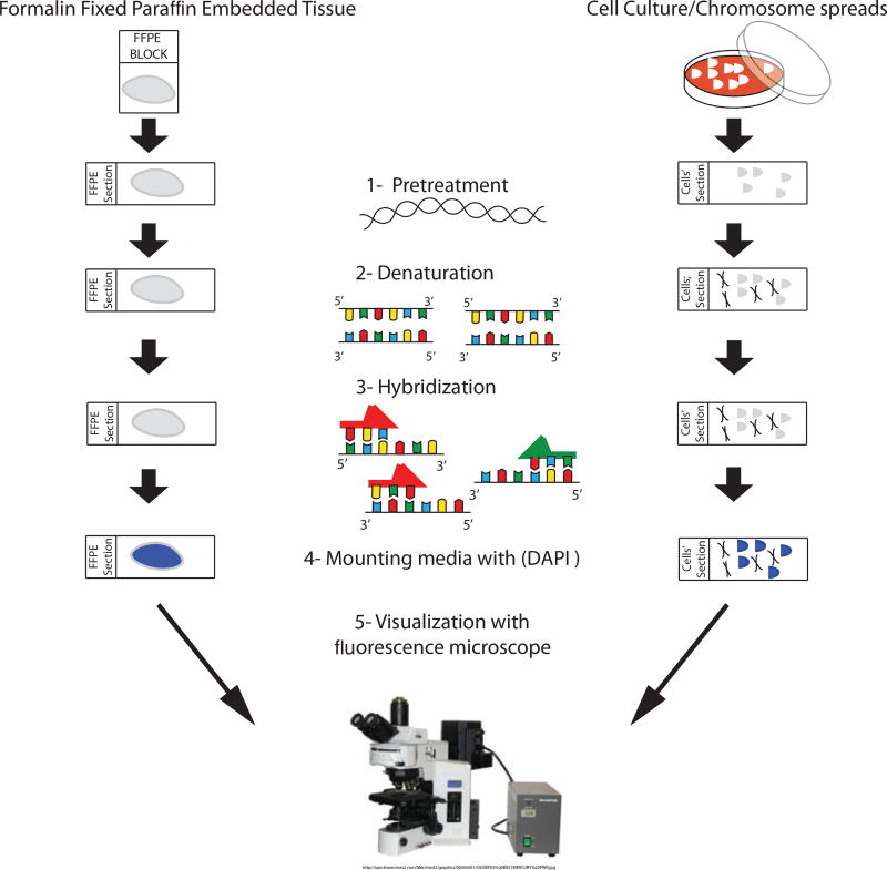
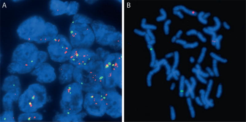
Fluorescence in situ hybridization (FISH) with DNA probes allows the visualization of gene copy number and localization of specific DNA targets with fluorescence microscopy. Cells in culture, metaphase chromosomes, and tissue sections are fixed and prepared on glass slides. Both the DNA in the cells and fluorescently labeled probe are denatured, and the labeled probe is allowed to hybridize to the cellular DNA. The slides are washed, counterstained, and viewed via fluorescence microscopy. We describe the basic method for preparing slides and probes for studies involving DNA copy number changes and structural chromosome rearrangements in formalin-fixed paraffin-embedded (FFPE) tissue sections and cell culture preparations.
Keywords: DNA probes; Fluorescence microscopy; Formalin-fixed paraffin-embedded tissues; Metaphase chromosomes.
Figures
References
-
- Kallioniemi A, Visakorpi T, Karhu R, Pinkel D, Kallioniemi OP. Gene copy number analysis by fluorescence in situ hybridization and comparative genomic hybridization. Methods. 1996;9(1):113–121. - PubMed
-
- Pinkel D, Gray JW, Trask B, van den Engh G, Fuscoe J, van Dekken H. Cytogenetic analysis by in situ hybridization with fluorescently labeled nucleic acid probes. Cold Spring Harb Symp Quant Biol. 1986;51(Pt 1):151–157. - PubMed
-
- Cajulis RS, Frias-Hidvegi D, Yu GH, Eggena S. Detection of numerical chromosomal abnormalities by fluorescence in situ hybridization of interphase cell nuclei with chromosome-specific probes on archival cytologic samples. Diagn Cytopathol. 1996;14(2):178–181. doi: 10.1002/(SICI)1097-0339(199603)14:2<178::AID--DC14>3.0.CO;2-J. - DOI - PubMed
-
- Cremer T, Lichter P, Borden J, Ward DC, Manuelidis L. Detection of chromosome aberrations in metaphase and interphase tumor cells by in situ hybridization using chromosome-specific library probes. Hum Genet. 1988;80(3):235–246. - PubMed
MeSH terms
Substances
Grants and funding
LinkOut - more resources
Full Text Sources
Other Literature Sources
