Multicenter Study
. 2018 Apr;55(4):3477-3489.
doi: 10.1007/s12035-017-0535-1. Epub 2017 May 13.

A Clinical and Molecular Genetic Study of 50 Families with Autosomal Recessive Parkinsonism Revealed Known and Novel Gene Mutations

Affiliations
Multicenter Study

A Clinical and Molecular Genetic Study of 50 Families with Autosomal Recessive Parkinsonism Revealed Known and Novel Gene Mutations

Shaghayegh Taghavi et al. Mol Neurobiol. 2018 Apr.

Abstract

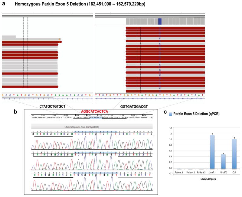
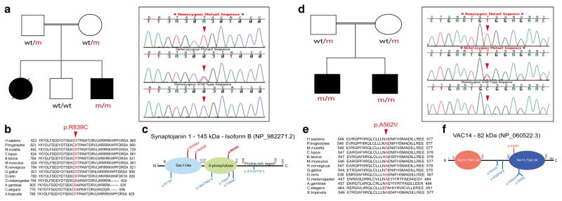
In this study, the role of known Parkinson's disease (PD) genes was examined in families with autosomal recessive (AR) parkinsonism to assist with the differential diagnosis of PD. Some families without mutations in known genes were also subject to whole genome sequencing with the objective to identify novel parkinsonism-related genes. Families were selected from 4000 clinical files of patients with PD or parkinsonism. AR inheritance pattern, consanguinity, and a minimum of two affected individuals per family were used as inclusion criteria. For disease gene/mutation identification, multiplex ligation-dependent probe amplification, quantitative PCR, linkage, and Sanger and whole genome sequencing assays were carried out. A total of 116 patients (50 families) were examined. Fifty-four patients (46.55%; 22 families) were found to carry pathogenic mutations in known genes while a novel gene, not previously associated with parkinsonism, was found mutated in a single family (2 patients). Pathogenic mutations, including missense, nonsense, frameshift, and exon rearrangements, were found in Parkin, PINK1, DJ-1, SYNJ1, and VAC14 genes. In conclusion, variable phenotypic expressivity was seen across all families.

Keywords: Early-onset; Genotype-phenotype correlations; Parkinson’s disease; Pathogenic mutations.


Conflict of interest statement

Conflict of Interest The authors declare that they have no competing interests.

Figures

Fig. 1
Fig. 1
Parkin exon 5 deletion (128-kb). a WGS reads were visualized by using the Integrative Genomics Viewer (IGV). Two different plots that represent the Parkin exon 5 deletion breakpoints are shown. b Sanger chromatograms of the three affected individuals, who carried the 128-kb Parkin exon 5 deletion, are shown. Deletion breakpoints are highlighted in black (12 bp of the flanking regions), while the 12-bp insertion identified within the deletion breakpoints is highlighted in red. c Validation of the Parkin exon 5 deletion through qPCR analyses. Y-axis represents PRKN/GPR15 ratios; x-axis: DNA samples analyzed. Affected individuals showed no copy of Parkin exon 5 (ratio = 0.0), whereas unaffected individuals showed either two copies (Unaff 1; ratio = 1.0–1.2) or one copy (Unaff 2; ratio = 0.4–0.6). Unaff unaffected family member, Ctrl control DNA sample (color figure online)
Fig. 2
Fig. 2
SYNJ1 p.Arg839Cys and VAC14 p.Ala562Val mutations. a Pedigree structure of the family carrying the SYNJ1 p.R839C mutation is shown on the left side. Affected family members are represented with black circle (female) or square (male). Wt/m heterozygous mutation carriers; m/m homozygous mutation carriers; wt/wt non-carriers. Sanger chromatogram sequences belonging to the SYNJ1 p.R839C mutation (red arrow) are shown on the right side. b Conservation of the SYNJ1 p.R839C mutation across different orthologs. c Diagram of the Synaptojanin 1 protein structure. The three SYNJ1 mutations identified to date in patients with parkinsonism are represented in red, whereas the SYNJ1 mutations identified in patients with seizures and severe neurodegeneration are shown in blue. The mutation identified in the current study is highlighted in bold. d Pedigree structure of the family carrying the VAC14 p.A562V mutation is shown on the left side. Affected family members are represented with black squares (males). Wt/m heterozygous mutation carriers; m/m homozygous mutation carriers; wt/wt non-carriers. Sanger chromatogram sequences for the VAC14 p.A562V mutation (red arrow) are shown on the right side. e Conservation of the VAC14 p.A562V mutation across different species. f Diagram of the Vac14 protein structure showing the mutation identified in patients with dystonic tremor and disabling dystonia at the top (bold) and the mutations identified in patients with striatonigral neurodegeneration at the bottom

References

    1. Tysnes OB, Storstein A. Epidemiology of Parkinson’s disease. J Neural Transm (Vienna) 2017 doi: 10.1007/s00702-017-1686-y. - DOI - PubMed
    1. Surmeier DJ, Obeso JA, Halliday GM. Selective neuronal vulnerability in Parkinson disease. Nat Rev Neurosci. 2017;18(2):101–113. doi: 10.1038/nrn.2016.178. - DOI - PMC - PubMed
    1. Halliday G, Lees A, Stern M. Milestones in Parkinson’s disease—clinical and pathologic features. Mov Disord. 2011;26(6):1015–1021. doi: 10.1002/mds.23669. - DOI - PubMed
    1. Lees AJ, Hardy J, Revesz T. Parkinson’s disease. Lancet. 2009;373(9680):2055–2066. doi: 10.1016/S0140-6736(09)60492-X. - DOI - PubMed
    1. Hardy J, Lewis P, Revesz T, Lees A, Paisan-Ruiz C. The genetics of Parkinson’s syndromes: a critical review. Curr Opin Genet Dev. 2009;19(3):254–265. - PubMed

Publication types