. 2017 Jun 5;14(6):1852-1860.
doi: 10.1021/acs.molpharmaceut.6b01015. Epub 2017 May 19.

Leveraging Colloidal Aggregation for Drug-Rich Nanoparticle Formulations

Affiliations

Leveraging Colloidal Aggregation for Drug-Rich Nanoparticle Formulations

Ahil N Ganesh et al. Mol Pharm. .

Abstract

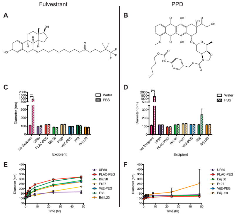
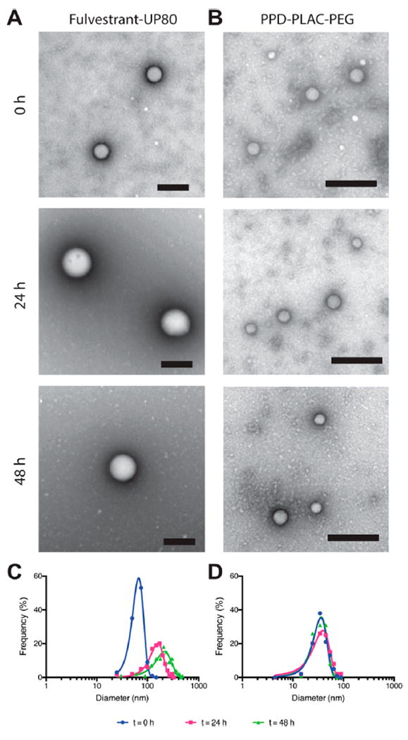
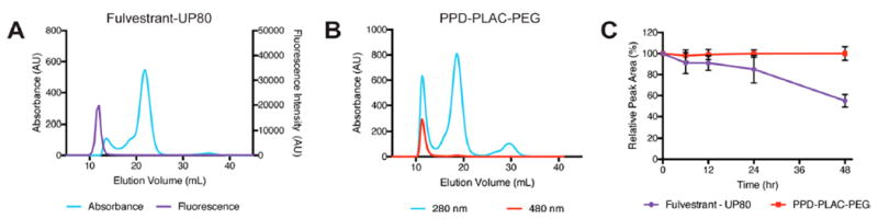
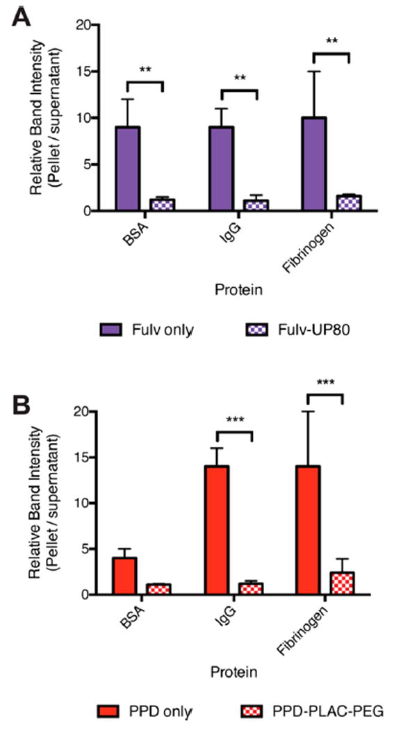
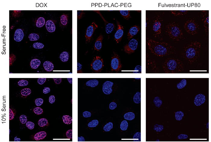
While limited drug loading continues to be problematic for chemotherapeutics formulated in nanoparticles, we found that we could take advantage of colloidal drug aggregation to achieve high loading when combined with polymeric excipients. We demonstrate this approach with two drugs, fulvestrant and pentyl-PABC doxazolidine (PPD; a prodrug of doxazolidine, which is a DNA cross-linking anthracycline), and two polymers, polysorbate 80 (UP80) and poly(d,l-lactide-co-2-methyl-2-carboxytrimethylene carbonate)-graft-poly(ethylene glycol) (PLAC-PEG; a custom-synthesized, self-assembling amphiphilic polymer). In both systems, drug-loaded nanoparticles had diameters < 200 nm and were stable for up to two days in buffered saline solution and for up to 24 h in serum-containing media at 37 °C. While colloidal drug aggregates alone are typically unstable in saline and serum-containing media, we attribute the colloid stability observed herein to the polymeric excipients and consequent decreased protein adsorption. We expect this strategy of polymer-stabilized colloidal drug aggregates to be broadly applicable in delivery formulations.

Keywords: colloids; drug delivery; polymers; self-assembly; solvent exchange.


Conflict of interest statement

Notes

The authors declare no competing financial interest.

Figures

Figure 1
Figure 1
(A) Fulvestrant and (B) PPD were selected for their intrinsic chemotherapeutic efficacy and aggregation properties. Formulation of (C) fulvestrant and (D) PPD colloids in water or PBS in the presence of the following polymeric excipients: UP80, PLAC–PEG, Brij 58, Pluronic F127, VitE-PEG, Pluronic F68, and Brij L23. Initial diameters of the formulations are shown. Incubation of (E) fulvestrant and (F) PPD formulations at 37 °C, showing size changes over 48 h. UP80 was the optimal polymer to maintain the size of fulvestrant over time in buffered salt solution (PBS) compared to other polymers. Stability of PPD with F68 could not be assessed due to precipitation. PLAC–PEG was the optimal polymer to maintain the size of PPD, with the smallest nanoparticle size over the incubation period. (n = 3, mean + SD, *** p < 0.001.)
Figure 2
Figure 2
Fulvestrant–UP80 and PPD–PLAC–PEG characterization in 10% serum. (A, B) Representative TEM images of particles in serum at 0, 24, and 48 h. (A) Fulvestrant–UP80 colloids were stained with ammonium molybdate, and (B) PPD–PLAC–PEG colloids were stained with uranyl acetate; scale bars are 200 nm. (C, D) TEM frequency distribution showing a mean diameter increase and peak broadening of (C) fulvestrant–UP80 colloidal aggregates over time, whereas (D) PPD–PLAC–PEG maintains its size and distribution over time.
Figure 3
Figure 3
Serum stability of colloidal formulations by FPLC. Traces show separation between serum proteins (absorbance at 280 nm) and (A) fulvestrant–UP80 colloids (tracked by fluorescence using a BODIPY FRET pair) and (B) PPD–PLAC–PEG colloids (unique absorbance at 480 nm) at t = 0. (C) Comparison of the peak area under the colloid curve over time to the area at t = 0 h to determine colloid stability as a function of time. Both colloids are stable up to 24 h, with fulvestrant–UP80 colloids dissociating between 24 and 48 h and PPD–PLAC–PEG colloids showing no evidence of dissociation over 48 h.
Figure 4
Figure 4
Formulation of colloids with excipient polymers reduces protein adsorption. BSA, IgG, and fibrinogen (50 nM) adsorption are significantly increased on the surface of bare colloids of (A) fulvestrant and (B) PPD (filled bars) compared to colloids stabilized with the appropriate polymer (checkered bars): fulvestrant–UP80 and PPD–PLAC–PEG (n = 3, mean + SD, **p < 0.01, ***p < 0.001).
Figure 5
Figure 5
Representative images of the cell uptake of DOX (monomer) and the colloidal formulations of PPD–PLAC–PEG and fulvestrant–UP80 (tracked by BODIPY). SKOV-3 cells were used for all experiments. DOX monomer freely permeates the cell membrane, showing localized fluorescence within the cells’ nuclei. PPD and fulvestrant colloids show uptake only under serum-free conditions, with punctate fluorescence within the cell body. There is no evidence of cell uptake of colloids in serum-containing media. (Scale bar represents 30 μm.)

References

    1. Strickley RG. Solubilizing Excipients in Oral and Injectable Formulations. Pharm Res. 2004;21(2):201–230. - PubMed
    1. Kiss L, Walter FR, Bocsik A, Veszelka S, Ózsvári B, Puskás LG, Szabó-Révész P, Deli MA. Kinetic Analysis of the Toxicity of Pharmaceutical Excipients Cremophor EL and RH40 on Endothelial and Epithelial Cells. J Pharm Sci. 2013;102(4):1173–1181. - PubMed
    1. Bravo Gonzalez RC, Huwyler J, Boess F, Walter I, Bittner B. In Vitro Investigation on the Impact of the Surface-Active Excipients Cremophor EL, Tween 80 and Solutol HS 15 on the Metabolism of Midazolam. Biopharm Drug Dispos. 2004;25(1):37–49. - PubMed
    1. Peer D, Karp JM, Hong S, Farokhzad OC, Margalit R, Langer R. Nanocarriers as an Emerging Platform for Cancer Therapy. Nat Nanotechnol. 2007;2(12):751–760. - PubMed
    1. Cabral H, Kataoka K. Progress of Drug-Loaded Polymeric Micelles Into Clinical Studies. J Controlled Release. 2014;190:465–476. - PubMed

Publication types