. 2017 Jul;187(7):1485-1495.
doi: 10.1016/j.ajpath.2017.03.010. Epub 2017 May 11.

Cyst-Like Osteolytic Formations in Recombinant Human Bone Morphogenetic Protein-2 (rhBMP-2) Augmented Sheep Spinal Fusion

Affiliations

Cyst-Like Osteolytic Formations in Recombinant Human Bone Morphogenetic Protein-2 (rhBMP-2) Augmented Sheep Spinal Fusion

Hsin Chuan Pan et al. Am J Pathol. 2017 Jul.

Abstract

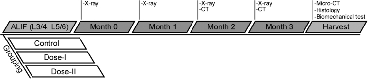
Multiple case reports using recombinant human bone morphogenetic protein-2 (rhBMP-2) have reported complications. However, the local adverse effects of rhBMP-2 application are not well documented. In this report we show that, in addition to promoting lumbar spinal fusion through potent osteogenic effects, rhBMP-2 augmentation promotes local cyst-like osteolytic formations in sheep trabecular bones that have undergone anterior lumbar interbody fusion. Three months after operation, conventional computed tomography showed that the trabecular bones of the rhBMP-2 application groups could fuse, whereas no fusion was observed in the control group. Micro-computed tomography analysis revealed that the core implant area's bone volume fraction and bone mineral density increased proportionately with rhBMP-2 dose. Multiple cyst-like bone voids were observed in peri-implant areas when using rhBMP-2 applications, and these sites showed significant bone mineral density decreases in relation to the unaffected regions. Biomechanically, these areas decreased in strength by 32% in comparison with noncystic areas. Histologically, rhBMP-2-affected void sites had an increased amount of fatty marrow, thinner trabecular bones, and significantly more adiponectin- and cathepsin K-positive cells. Despite promoting successful fusion, rhBMP-2 use in clinical applications may result in local adverse structural alterations and compromised biomechanical changes to the bone.


Figures

Figure 1
Figure 1
Experimental design. Spinal fusion (ALIF) operation was performed on animals randomly assigned to three different groupings. The analysis after the operation included monthly X-ray scans and conventional CT scans at months 2 and 3. At 3 months after the operation, samples were harvested and underwent high-resolution micro-CT, biomechanical, and histologic analyses. ALIF, anterior lumbar interbody fusion; CT, computed tomography; Dose-I, 0.43 mg/mL (0.65 mg in total) recombinant human bone morphogenetic protein-2 and an absorbable collagen sponge in the spacer; Dose-II, 1.5 mg/mL (2.25 mg in total) recombinant human bone morphogenetic protein-2 and an absorbable collagen sponge in the spacer. n = 4.
Figure 2
Figure 2
Results of conventional CT. A and B: Representative conventional CT images from each group 2 (A) and 3 (B) months after the operation. Successful fusion was observed in all rhBMP-2 groups, and no union was observed in the control group. The bone volume fraction within the spacer was calculated by measuring the ratio of new bone volume against the entire spacer volume at five vertical levels that were evenly distanced when positioned from an axial view. Volumetric deformity was defined as the bone volume fraction occupying <90% of the spacer. In an axial view, a volumetric deformity of the implant core (arrow) was observed in the dose-I group at both time points. Scale bar = 5 mm. CT, computed tomography; Dose-I, 0.43 mg/mL (0.65 mg in total) rhBMP-2 and an absorbable collagen sponge in the spacer; Dose-II, 1.5 mg/mL (2.25 mg in total) rhBMP-2 and an absorbable collagen sponge in the spacer; rhBMP-2, recombinant human bone morphogenetic protein-2.
Figure 3
Figure 3
3D orthogonal slice representatives from high-resolution micro-CT. A: The control group presented with a clear bony gap in all samples. Both rhBMP-2 groups showed solid fusion, but the dose-I group revealed a volumetric deformity toward the center. B: Axial view slice selection was based on the midpoint of the implant core's vertical dimension. Sagittal and coronal slices were determined from the center of the implant. Scale bar = 5 mm. 3D, three-dimensional; CT, computed tomography; Dose-I, 0.43 mg/mL (0.65 mg in total) rhBMP-2 and an absorbable collagen sponge in the spacer; Dose-II, 1.5 mg/mL (2.25 mg in total) rhBMP-2 and an absorbable collagen sponge in the spacer; rhBMP-2, recombinant human bone morphogenetic protein-2.
Figure 4
Figure 4
Quantitative analysis of fusion mass with high-resolution micro-CT. Fusion quality was quantitatively analyzed by dividing the sample into the core and peri-implant areas. A: The core area showed dosage-dependent BMD and BV/TV increases. B: The peri-implant area shows that the BMD was significantly lower in the dose-I group than in the control group. C: Core implant VOI-1 was defined as a cylindrical volume of 7 mm in height and diameter in the center of the spacer. Peri-implant VOI-2 was defined as the volume directly above and below the implant spacer, and following the spacer contour. The volume height was 7 mm, because this was the furthest distance after comparing all samples in which cyst-like bone voids were observed. P < 0.05, ∗∗P < 0.01. BMD, bone mineral density; BV/TV, bone volume/tissue volume; CT, computed tomography; Dose-I, 0.43 mg/mL (0.65 mg in total) recombinant human bone morphogenetic protein-2 and an absorbable collagen sponge in the spacer; Dose-II, 1.5 mg/mL (2.25 mg in total) recombinant human bone morphogenetic protein-2 and an absorbable collagen sponge in the spacer; Tb.Th, trabecular thickness; VOI, volume of interest.
Figure 5
Figure 5
Representative images of cyst-like bone voids and quantitative analysis. From a macro to micro scale, the cyst-like bone void sites were analyzed. A: For the control, no cystic change was found throughout the sample. B and C: The dose-I group shows small clustered bone cysts over an area, whereas the dose-II group sample could localize as a solitary lesion. A 27-mm3 VOI-3 was selected after reviewing all of the cyst-like bone void regions and was analyzed quantitatively to investigate the relative density/volume change. Orange boxed areas are locations where subsequent micro-CT illustrations (shown below) were extracted from, with white boxed areas depicting the 27-mm3 cubic volumes used for analysis. D: The cubic volume was selected because of its relative position to the implant core, with one cubic volume directly above it and another more distant, but still adjacent to the spacer. The cyst-like bone void area showed significant bone weakness (low BMD and BV/TV) compared with the unaffected sites. P < 0.05, ∗∗P < 0.01. Scale bar = 3 mm. BMD, bone mineral density; BV/TV, bone volume/tissue volume; CT, computed tomography; Dose-I, 0.43 mg/mL (0.65 mg in total) recombinant human bone morphogenetic protein-2 and an absorbable collagen sponge in the spacer; Dose-II, 1.5 mg/mL (2.25 mg in total) recombinant human bone morphogenetic protein-2 and an absorbable collagen sponge in the spacer; VOI, volume of interest.
Figure 6
Figure 6
Biomechanical testing using FEA. A uniform compressive force of 0.5 MPa was applied to the superior surface of the 27-mm3 cubic-shaped VOI of both noncystic and cyst-like bone void areas. A: Cuboidal specimens from cyst-like bone void area showed an increased von Mises stress with higher intensity of colors (gray regions indicate all values exceeding 25 MPa). B: Quantification of cuboidal segments of each sample bone demonstrated significantly increased von Mises stress in the cyst-like bone void, which suggested deteriorated bone strength. P < 0.05. Scale bar = 3 mm. Dose-I, 0.43 mg/mL (0.65 mg in total) recombinant human bone morphogenetic protein-2 and an absorbable collagen sponge in the spacer; Dose-II, 1.5 mg/mL (2.25 mg in total) recombinant human bone morphogenetic protein-2 and an absorbable collagen sponge in the spacer; FEA, finite element analysis; S, Mises, von Mises stress; SNEG, surface negative; VOI, volume of interest.
Figure 7
Figure 7
Histologic examination of cyst-like bone voids. A–C: The cyst-like bone voids were located immediately next to rhBMP-2 implants in both treatment groups as opposed to the control (asterisks). D–F: The expanded spaces that had fewer and thinner trabecular bones were seen in the cyst-like bone voids but not in the control sample. G–I: A profound amount of fatty marrow filled the cyst-like bone voids in both rhBMP-2 samples. D–F are magnified views from asterisk areas in A–C. The boxed areas in D–F are shown below at higher magnification (G–I). Scale bars: 1 mm (top row); 200 μm (middle row); 100 μm (bottom row). Dose-I, 0.43 mg/mL (0.65 mg in total) rhBMP-2 and an absorbable collagen sponge in the spacer; Dose-II, 1.5 mg/mL (2.25 mg in total) rhBMP-2 and an absorbable collagen sponge in the spacer; H&E, hematoxylin and eosin; rhBMP-2, recombinant human bone morphogenetic protein-2.
Figure 8
Figure 8
Immunohistochemistry of adiponectin and cathepsin K. A–C: Many more adiponectin-positive cells present in the marrow cavity were detected in the rhBMP-2 groups (arrows) than in the control sample (arrow). D–F: An increased number of cathepsin K-positive osteoclasts along trabecular bones and in marrow cavity were detected in the rhBMP2 groups (arrows) than in the control group (arrow). Scale bar = 100 μm. Dose-I, 0.43 mg/mL (0.65 mg in total) rhBMP-2 and an absorbable collagen sponge in the spacer; Dose-II, 1.5 mg/mL (2.25 mg in total) rhBMP-2 and an absorbable collagen sponge in the spacer; rhBMP-2, recombinant human bone morphogenetic protein-2.
Supplemental Figure S1
X-ray of the lumbar spine immediately after the operation. The implant position was confirmed after the operation with anteroposterior and lateral X-ray radiographs. The subsequent positioning and fusion assessments took place immediately after the operation and 1, 2, and 3 months after the operation. Vertebral spacers are observed at both L3-L4 and L5-L6 levels of each group. Scale bar = 3 cm. AP, anteroposterior; Dose-I, 0.43 mg/mL (0.65 mg in total) recombinant human bone morphogenetic protein-2 and an absorbable collagen sponge in the spacer; Dose-II, 1.5 mg/mL (2.25 mg in total) recombinant human bone morphogenetic protein-2 and an absorbable collagen sponge in the spacer.
Supplemental Figure S2
Additional histology and immunohistochemistry on cyst-like bone voids. A: The inflammatory cells and a dilated blood vessel (arrow) and a piece of dead bone (arrowhead) were observed in the rhBMP-2 sample. B: Adiponectin-positive staining pattern on mature fatty drop-filled adipocytes (arrows). C: The negative staining control generated by replacing specific adiponectin or cathepsin K antibodies with PBS on a rhBMP-2 sample. Scale bar = 100 μm. Dose-I, 0.43 mg/mL (0.65 mg in total) rhBMP-2 and an absorbable collagen sponge in the spacer; H&E, hematoxylin and eosin; PBS, phosphate-buffered saline; rhBMP-2, recombinant human bone morphogenetic protein-2.
Supplemental Figure S3
Rat posterolateral spinal fusion using rhBMP-2. As the preliminary study, posterolateral spinal fusions were performed using two different doses of rhBMP-2: 100 μg (A) and 200 μg (B) treatment group. Samples from the rats were harvested 3 weeks after the operation. Although both groups have successful implant fusions, as observed in the 3D reconstructed images, large volumes of cyst-like bone voids (arrow) are apparent in the orthogonal sectional images. As a result, the implant fusions are not completely solid. Scale bar = 3 mm. CT, computed tomography; rhBMP-2, recombinant human bone morphogenetic protein-2; 3D, three-dimensional.

References

    1. Burkus J.K., Sandhu H.S., Gornet M.F., Longley M.C. Use of rhBMP-2 in combination with structural cortical allografts: clinical and radiographic outcomes in anterior lumbar spinal surgery. J Bone Joint Surg Am. 2005;87:1205–1212. - PubMed
    1. Mesfin A., Buchowski J.M., Zebala L.P., Bakhsh W.R., Aronson A.B., Fogelson J.L., Hershman S., Kim H.J., Ahmad A., Bridwell K.H. High-dose rhBMP-2 for adults: major and minor complications: a study of 502 spine cases. J Bone Joint Surg Am. 2013;95:1546–1553. - PubMed
    1. Knox J.B., Dai J.M., III, Orchowski J. Osteolysis in transforaminal lumbar interbody fusion with bone morphogenetic protein-2. Spine (Phila Pa 1976) 2011;36:672–676. - PubMed
    1. Garrett M.P., Kakarla U.K., Porter R.W., Sonntag V.K. Formation of painful seroma and edema after the use of recombinant human bone morphogenetic protein-2 in posterolateral lumbar spine fusions. Neurosurgery. 2010;66:1044–1049. discussion 1049. - PubMed
    1. McClellan J.W., Mulconrey D.S., Forbes R.J., Fullmer N. Vertebral bone resorption after transforaminal lumbar interbody fusion with bone morphogenetic protein (rhBMP-2) J Spinal Disord Tech. 2006;19:483–486. - PubMed

MeSH terms

Substances