. 2016 Oct;9(10):238-247.
doi: 10.4172/jpb.1000412. Epub 2016 Oct 3.

Protein Markers Associated with an ALDH Sub-Population in Colorectal Cancer

Affiliations

Protein Markers Associated with an ALDH Sub-Population in Colorectal Cancer

Rui Yang et al. J Proteomics Bioinform. 2016 Oct.

Abstract

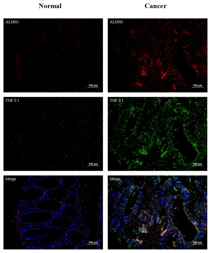
ALDH has been shown to be a marker that denotes a sub-population of cancer stem cells in colorectal and other cancers. This sub-population of cells shows an increased risk for tumor initiation, metastasis, and resistance to chemotherapy and radiation resulting in recurrence and death. It is thus essential to identify the important signaling pathways related to ALDH1+ CSCs in colon cancer. The essential issue becomes to isolate pure sub-populations of cells from heterogeneous tissues for further analysis. To achieve this goal, tissues from colorectal cancer Stage III patients were immuno-stained with ALDH1 antibody. Target ALDH1+ and ALDH1- cells from the same tissue were micro-dissected using Laser Capture Microdissection (LCM). Captured cells were lysed and analyzed using LC-MS/MS where around 20,000 cells were available for analysis. This analysis resulted in 134 proteins which were differentially expressed between ALDH1+ and ALDH1- cells in three patient sample pairs. Based on these differentially expressed proteins an IPA pathway analysis was performed that showed two key pathways in cell to cell signaling and organismal injury and abnormalities. The IPA analysis revealed β-catenin, NFκB (p65) and TGFβ1 as important cancer-related proteins in these pathways. A TMA validation using immunofluorescence staining of tissue micro-arrays including 170 cases was used to verify that these key proteins were highly overexpressed in ALDH1+ cells in colon cancer tissues compared to ALDH1- cells.

Keywords: ALDH; Colon cancer; FFPE tissue section; LC-MS/MS; LCM.


Figures

Figure 1
Figure 1
The proteomic workflow used in this study is summarized. We have developed a method of analyzing pure cell subpopulations isolated from FFPE tissue sections combining immunohistochemistry, laser microdissection, and shotgun proteome analysis.
Figure 2
Figure 2
Canonical signaling pathways enriched with commonly differentially expressed proteins between ALDH1+ and ALDH1− cells and ranked by significance in the IPA. A p-value threshold of 0.01 is applied.
Figure 3
Figure 3
Top two connectivity networks constructed by IPA: (A) Cell-to-Cell Signaling; (B) Organismal Injury and Abnormalities. These networks only consist of differentially expressed proteins derived from the experimental data. Red and green represent over- and under-expression in ALDH1+ cells compared with ALDH1- cells, respectively. White indicates proteins that were not identified as differentially expressed in this study or were not detected by the mass spec analysis but are relevant to the networks.
Figure 4
Figure 4
Immunofluorescence double staining with ALDH1 (red) and β-catenin (green) in human normal colon tissue and colon cancer. DAPI represent the nucleus of the cells. Overall, the expressions of ALDH1 and β-catenin are negative in normal tissues. ALDH1 is positive on cell membranes. However, both of them are overexpressed in cancer tissues. β-catenin shows strong nucleus positive staining and is highly overlapped with ALDH1. Magnification 200x, scale bars=100 μm.
Figure 5
Figure 5
Immunofluorescence double staining with ALDH1 (green) and NFκB(p65) (red) in human normal colon tissue and colon cancer. DAPI represent the nucleus of the cells. There is no expression of ALDH1 and NFκB(p65) in normal tissues. NFκB(p65) is highly expressed in the cytoplasm. The overlaps have been shown in the merged images. Magnification 200x, scale bars=100 μm.
Figure 6
Figure 6
Immunofluorescence double staining with ALDH1 (red) and TGFβ1 (green) in human normal colon tissue and colon cancer. DAPI staining represents the nucleus of the cells. The expressions of ALDH1 and TGFβ1 are significantly increased in cancer tissues compared to normal tissues. TGFβ1 shows strong cell membranes and extracellular positive staining. Magnification 200x, scale bars=100 μm.
Figure 7
Figure 7
Association between ALDH1+ & β-catenin+ overlap expression and stages. X axis represents different stages. And Y axis represents the percentages of the overlaps. All overlap cases (n=92) and the medians are shown in this figure.

References

    1. Edwards TL, Shrubsole MJ, Cai Q, Li G, Dai Q, et al. A study of prostaglandin pathway genes and interactions with current nonsteroidal anti-inflammatory drug use in colorectal adenoma. Cancer Prev Res. 2012;5:855–863. - PMC - PubMed
    1. Escoffery C, Fernandez ME, Vernon SW, Liang S, Maxwel AE, et al. Patient Navigation in a Colorectal Cancer Screening Program. J Public Health Manag Pract. 2015;21:433–440. - PMC - PubMed
    1. Weyl H, Yackzan S, Ross K, Henson A, Moe K, et al. Understanding colorectal screening behaviors and factors associated with screening in a community hospital setting. Clin J Oncol Nurs. 2015;19:89–93. - PubMed
    1. Sabbagh C, Chatelain D, Trouillet N, Mauvais F, Bendjaballah S, et al. Does use of a metallic colon stent as a bridge to surgery modify the pathology data in patients with colonic obstruction? A case-matched study. Surg Endosc. 2013;27:3622–3631. - PubMed
    1. Lurje G, Zhang W, Lenz HJ. Molecular prognostic markers in locally advanced colon cancer. Clin Colorectal Cancer. 2007;6:683–690. - PubMed

LinkOut - more resources