Review
. 2017 Apr 28:8:177.
doi: 10.3389/fneur.2017.00177. eCollection 2017.

Superior Canal Dehiscence Syndrome: Lessons from the First 20 Years

Affiliations
Review

Superior Canal Dehiscence Syndrome: Lessons from the First 20 Years

Bryan K Ward et al. Front Neurol. .

Abstract

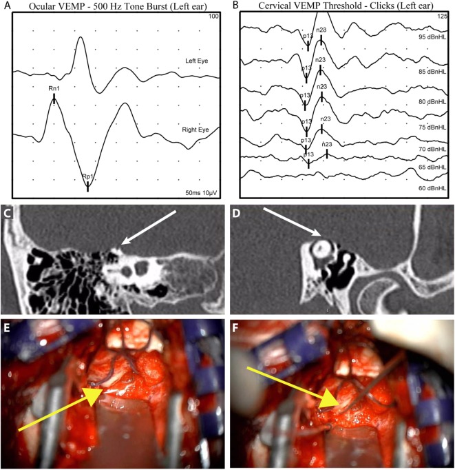
Superior semicircular canal dehiscence syndrome was first reported by Lloyd Minor and colleagues in 1998. Patients with a dehiscence in the bone overlying the superior semicircular canal experience symptoms of pressure or sound-induced vertigo, bone conduction hyperacusis, and pulsatile tinnitus. The initial series of patients were diagnosed based on common symptoms, a physical examination finding of eye movements in the plane of the superior semicircular canal when ear canal pressure or loud tones were applied to the ear, and high-resolution computed tomography imaging demonstrating a dehiscence in the bone over the superior semicircular canal. Research productivity directed at understanding better methods for diagnosing and treating this condition has substantially increased over the last two decades. We now have a sound understanding of the pathophysiology of third mobile window syndromes, higher resolution imaging protocols, and several sensitive and specific diagnostic tests. Furthermore, we have a treatment (surgical occlusion of the superior semicircular canal) that has demonstrated efficacy. This review will highlight some of the fundamental insights gained in SCDS, propose diagnostic criteria, and discuss future research directions.

Keywords: autophony; labyrinth diseases; superior semicircular canal dehiscence syndrome; vertigo; vestibular diseases.


Figures

Figure 1
Figure 1
Number of publications related to SCDS by year since its original description. Figures derived from PubMed search for “superior canal dehiscence.”
Figure 2
Figure 2
Original graphical demonstrations by Cawthorne of the third mobile window effect. (A) Demonstrates the effect of semicircular canal fenestration in otosclerosis with a fixed stapes footplate and two mobile windows and (B) third mobile window established via semicircular canal fenestration in a patient with a mobile stapes footplate.
Figure 3
Figure 3
Illustrative case to demonstrate diagnostic and intraoperative findings: a 40-year-old man presented with 6 years of left aural fullness, pulsatile tinnitus, vocal distortion, and hearing his eyeballs move in his left ear. Ocular vestibular-evoked myogenic potentials (VEMPs) indicated elevated amplitude responses to 500 Hz tone bursts [(A), 47.3 µV, normal range 0–17 µV] and cervical VEMPs with low thresholds in response to clicks [(B), 65 dB nHL, normal range ≥80 dB nHL], both suggestive of a third mobile window syndrome involving the left ear. High-resolution computed tomography imaging with 0.6-mm slice thickness demonstrated a dehiscence of the left superior semicircular canal when image reconstructions were made orthogonal to the plane of the superior canal [(C), Stenvers view] and in the plane of the superior canal [(D), Pöschl view]. He elected to proceed with surgery via middle cranial fossa approach. The dehiscence measured 5 mm × 1 mm [(E), yellow arrow] and was plugged with a combination of autologous materials including fascia, bone dust, and bone chips (F). The middle cranial fossa was resurfaced with hydroxyapatite cement. Autophony improved after surgery, hearing was preserved, and vestibular dysfunction was limited to the superior semicircular canal as determined by clinical head impulse testing in all semicircular canal planes.

References

    1. Minor LB, Solomon D, Zinreich JS, Zee DS. Sound- and/or pressure-induced vertigo due to bone dehiscence of the superior semicircular canal. Arch Otolaryngol Head Neck Surg (1998) 124:249–58.10.1001/archotol.124.3.249 - DOI - PubMed
    1. Minor LB. Superior canal dehiscence syndrome. Am J Otol (2000) 21:9–19.10.1016/S0196-0709(00)80105-2 - DOI - PubMed
    1. Rosowski JJ, Songer JE, Nakajima HH, Brinsko KM, Merchant SN. Clinical, experimental, and theoretical investigations of the effect of superior semicircular canal dehiscence on hearing mechanisms. Otol Neurotol (2004) 25:323–32.10.1097/00129492-200405000-00021 - DOI - PubMed
    1. Mikulec AA, McKenna MJ, Ramsey MJ, Rosowski JJ, Herrmann BS, Rauch SD, et al. Superior semicircular canal dehiscence presenting as conductive hearing loss without vertigo. Otol Neurotol (2004) 25:121–9.10.1097/00129492-200403000-00007 - DOI - PubMed
    1. Minor LB, Carey JP, Cremer PD, Lustig LR, Streubel S-O, Ruckenstein MJ. Dehiscence of bone overlying the superior canal as a cause of apparent conductive hearing loss. Otol Neurotol (2003) 24:270–8.10.1097/00129492-200303000-00023 - DOI - PubMed

LinkOut - more resources