. 2017 Apr;33(2):141-147.
doi: 10.5487/TR.2017.33.2.141. Epub 2017 Apr 15.

Spontaneous Peripheral Ameloblastic Odontoma in a Male Sprague-Dawley Rat

Affiliations

Spontaneous Peripheral Ameloblastic Odontoma in a Male Sprague-Dawley Rat

Yinghua Li et al. Toxicol Res. 2017 Apr.

Abstract

Peripheral ameloblastic odontoma is a rare variant of odontogenic tumor occurring in the extraosseous region. The present report describes a spontaneous tumor in male Sprague-Dawley (SD) rats. The clinically confirmed nodule in the right mandibular region was first observed when the rat was 42 weeks and remained until the terminal sacrifice date when the animal was 48 weeks of age. At necropsy, a well demarcated nodule, approximately 2.5 × 2.0 × 2.0 cm, protruded from the ventral area of the right mandible. The nodule was not attached to mandibular bone and was not continuous with the normal teeth. Histopathologically, the tumor was characterized by the simultaneous occurrence of an ameloblastomatous component and composite odontoma-like elements within the same tumor. The epithelial portion formed islands or cords resembling the follicle or plexiform pattern typical of ameloblastoma and was surrounded by mesenchymal tissue. Formation of eosinophilic and basophilic hard tissue matrix (dentin and enamel) resembling odontoma was observed in the center of the tumor. Mitotic figures were rare, and areas of cystic degeneration were present. Immunohistochemically, the epithelial component was positive for cytokeratin AE1/AE3 (CK AE1/AE3), and the mesenchymal component and odontoblast-like cells were positive for vimentin, in the same manner as in normal teeth. On the basis of these findings, the tumor was diagnosed as a peripheral ameloblastic odontoma in an extraosseous mandibular region in a SD rat. In the present study, we report the uncommon spontaneous peripheral ameloblastic odontoma in the SD rat. We also discuss here the morphological characteristics, origin, histochemical, and immunohistochemical features for the diagnosis of this tumor.

Keywords: Ameloblastoma; Odontogenic ectomesenchyme; Odontoma; Peripheral ameloblastic odontoma; Sprague-Dawley rat.


Figures

Fig. 1
Fig. 1
Gross appearance. The cut surface of the tumor appeared whitish after fixation. 1 scale = 0.5 cm.
Fig. 2
Fig. 2
(A) H&E staining. Odontogenic epithelial cells were arranged in cords or follicular patterns surrounded by mesenchymal cells. The ameloblast-like cells were palisaded in the peripheral area and the stellate reticulum-like cells were arranged in central area. Areas of cystic degeneration were present also (arrow). Scale bar = 200 μm. (B) H&E staining. The epithelial component contained eosinophilic granular cells at its center. Multinucleated polygonal cells were present along the surface of osseous tissue and eosinophilic material (arrows) was layered between the epithelium and the mesenchymal stroma. Scale bar = 50 μm. Inset: areas of squamous metaplasia surrounded by an ameloblast-like cells. (C) H&E staining. The abortive tooth structures consisted of a pulp cavity lined by a layer of ameloblast-like cells, dentin, and single layer of cuboidal to flattened odontoblast-like cells (arrows). Scale bar = 50 μm. (D) TRAP staining. The many TRAP-positive cells (purple cytoplasm) were multinucleated osteoclasts. Scale bar = 50 μm.
Fig. 3
Fig. 3
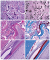
(A) H&E staining. The hard tissue matrix in the center of the tumor was basophilic when calcified, and eosinophilic when uncalcified. Scale bar = 200 μm. (B) von Kossa staining. Only hard tissue areas that were basophilic with H&E stain appeared positive for calcium (black) with von Kossa stain. Scale bar = 200 μm. (C) H&E staining. Abortive tooth-like structures and areas of ossification, osseous and squamous metaplasia (arrow) were present in the tumor. Scale bar = 50 μm. (D) Masson’s Trichrome staining. Eosinophilic matrix and ossification tissue (collagenous tissues) appeared blue with Masson’s trichrome stain. The deeply eosinophilic matrix, squamous metaplasia, and eosinophilic granular cells stained with H&E appeared dark red with Masson’s Trichrome stain. Scale bar = 50 μm. (E) H&E staining. Normal incisor tooth was appeared. Scale bar = 100 μm. (F) Masson‘s Trichrome staining. In normal teeth, dentin, bone, and fibrous tissue appeared blue and the enamel matrix appeared dark red. Scale bar = 100 μm.
Fig. 4
Fig. 4
Immunohistochemical staining. (A) The epithelial cell layer was severe positive for CK AE1/AE3. Scale bar = 100 μm. (B) The surrounding mesenchymal cells, odontoblast-like cells, and satellite reticulum-like cells were severe positive for vimentin. Scale bar = 100 μm. (C) In normal tooth, the ameloblasts were severe positive for the CK AE1/AE3. Scale bar = 100 μm. (D) In normal tooth, the dental pulp and odontoblasts were severe positive for vimentin. Scale bar = 100 μm.

Similar articles

Cited by

References

    1. Kramer IRH, Pindborg JJ, Shear M. Histological typing of odontogenic tumours. 2nd edition. Springer-Verlag Heidelberg; New York: 1992. World Health Organization (International histological classification of tumours) - DOI
    1. Burrough ER, Myers RK, Whitley EM. Spontaneous odontoameloblastoma in a female Sprague Dawley rat. J Vet Diagn Invest. 2010;22:998–1001. doi: 10.1177/104063871002200628. - DOI - PubMed
    1. Mosqueda-Taylor A, Carlos-Bregni R, Ramirez-Amador V, Palma-Guzman JM, Esquivel-Bonilla D, Hernandez-Rojase LA. Odontoameloblastoma. Clinic-pathologic study of three cases and critical review of the literature. Oral Oncol. 2002;38:800–805. doi: 10.1016/S1368-8375(02)00046-5. - DOI - PubMed
    1. Weber K. Induced and spontaneous lesions in teeth of laboratory animals. J Toxicol Pathol. 2007;20:203–213. doi: 10.1293/tox.20.203. - DOI
    1. Nold JB, Powers BE, Eden EL, McChesney AE. Ameloblastic odontoma in a dog. J Am Vet Med Assoc. 1984;185:996–998. - PubMed

LinkOut - more resources