Clinico- pathologic presentation of hypersensitivity pneumonitis in Egyptian patients: a multidisciplinary study
- PMID: 28503304
- PMCID: PMC5421323
- DOI: 10.1186/s40248-017-0091-6
Clinico- pathologic presentation of hypersensitivity pneumonitis in Egyptian patients: a multidisciplinary study
Abstract
Background: Hypersensitivity pneumonitis (HP) is a common diffuse parenchymal lung disease in Egypt which can be difficult to recognize due to the dynamic symptoms & associated environmental factors.
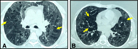
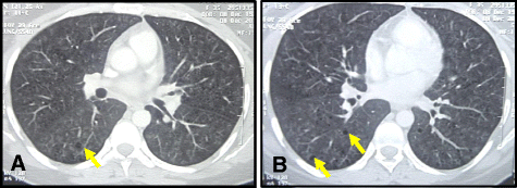
Methods: Forty-three Egyptian patients were enrolled in this study, presenting with dyspnea and cough, predominant ground-glass opacity (GGO) in high-resolution computed tomography (HRCT) where lung biopsy was needed to establish the diagnosis.
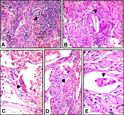
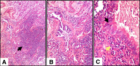
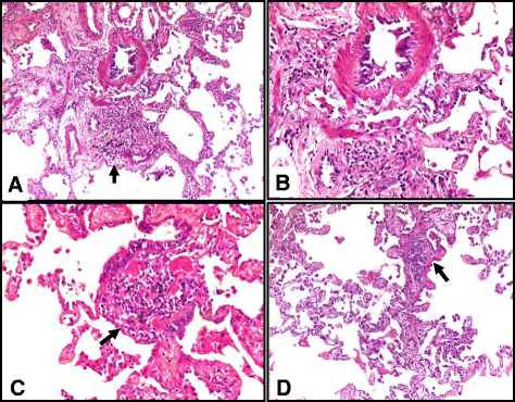
Results: The age range was 15 to 60 years. Females represented 90.7% (39 patients) while 9.3% (4 patients) of our patients were males. History of contact with birds was detected in 9 (20.9%) patients. Most of our patients (60.5%) didn't have exposure history, and only 8 patients (18.6%) were living in geographic areas in Egypt that are known for the exposure to environmental etiologic factors (cane sugar exhaust fumes). The most common HRCT pattern was GGO with mosaic parenchyma in 18 patients (41.86%), followed by GGO with centrilobular nodules in 9 patients (20.93%), then isolated diffuse GGO in 5 patients (11.62%), GGO with traction bronchiectasis in 4 patients (9.3%), GGO with consolidation in 3 patients (6.97%), GGO with reticulations in 2 patients (4.65%), and GGO with cysts in 2 patients (4.65%). The most common histologic finding was isolated multinucleated giant cells in 38 patients (88.3%) commonly found in airspaces (24 patients) and less commonly in the interstitium (14 patients), followed by interstitial pneumonia and cellular bronchiolitis in 36 patients (83.7% each), interstitial ill-formed non-necrotizing granulomas in 12 patients (27.9%), fibrosis in 10 patients (23.2%), and organizing pneumonia pattern in 4 patients (9.3%).
Conclusion: The diagnosis of HP presenting with predominant GGO pattern in HRCT requires a close interaction among clinicians, radiologists, and pathologists. Some environmental and household factors may be underestimated as etiologic factors. Further environmental and genetic studies are needed especially in patients with negative exposure history.
Keywords: Diffuse parenchymal lung diseases; Hypersensitivity pneumonitis; Interstitial lung diseases; Multidisciplinary approach.
Figures
Similar articles
-
The new useful high-resolution computed tomography finding for diagnosing fibrotic hypersensitivity pneumonitis: "hexagonal pattern": a single-center retrospective study.BMC Pulm Med. 2022 Mar 4;22(1):76. doi: 10.1186/s12890-022-01869-4. BMC Pulm Med. 2022. PMID: 35246090 Free PMC article.
-
Compatible with fibrotic hypersensitivity pneumonitis on high-resolution computed tomography: from the ATS/JRS/ALAT 2020 hypersensitivity pneumonitis guidelines.J Thorac Dis. 2024 Apr 30;16(4):2353-2364. doi: 10.21037/jtd-23-1845. Epub 2024 Apr 12. J Thorac Dis. 2024. PMID: 38738228 Free PMC article.
-
[Granulomatous diseases and pathogenic microorganism].Kekkaku. 2008 Feb;83(2):115-30. Kekkaku. 2008. PMID: 18326339 Japanese.
-
Hypersensitivity Pneumonitis: Essential Radiologic and Pathologic Findings.Surg Pathol Clin. 2010 Mar;3(1):187-98. doi: 10.1016/j.path.2010.03.005. Epub 2010 Jul 7. Surg Pathol Clin. 2010. PMID: 26839033 Review.
-
Clear vision through the haze: a practical approach to ground-glass opacity.Curr Probl Diagn Radiol. 2014 May-Jun;43(3):140-58. doi: 10.1067/j.cpradiol.2014.01.004. Curr Probl Diagn Radiol. 2014. PMID: 24791617 Review.
Cited by
-
Role of Krebs von den Lungen-6 (KL-6) in Assessing Hypersensitivity Pneumonitis.Tuberc Respir Dis (Seoul). 2021 Jul;84(3):200-208. doi: 10.4046/trd.2020.0122. Epub 2021 Apr 6. Tuberc Respir Dis (Seoul). 2021. PMID: 33840176 Free PMC article.
-
Krebs von den lungen-6 as a clinical marker for hypersensitivity pneumonitis: A meta-analysis and bioinformatics analysis.Front Immunol. 2022 Nov 30;13:1041098. doi: 10.3389/fimmu.2022.1041098. eCollection 2022. Front Immunol. 2022. PMID: 36532009 Free PMC article.
-
Study of nasal mucosa histopathological changes in patients with hypersensitivity pneumonitis.Sci Rep. 2023 May 31;13(1):8868. doi: 10.1038/s41598-023-35871-5. Sci Rep. 2023. PMID: 37258647 Free PMC article. Clinical Trial.
-
Predictors of pulmonary hypertension in patients with hypersensitivity pneumonitis.BMC Pulm Med. 2023 Feb 9;23(1):61. doi: 10.1186/s12890-023-02347-1. BMC Pulm Med. 2023. PMID: 36759788 Free PMC article. Clinical Trial.
References
-
- Madison JM. Hypersensitivity pneumonitis: clinical perspectives. Arch Pathol Lab Med. 2008;132(2):195–8. - PubMed
LinkOut - more resources
Full Text Sources
Other Literature Sources
Research Materials
Miscellaneous

