. 2014 Aug 26;1(1):2329048X14544955.
doi: 10.1177/2329048X14544955. eCollection 2014 Jul-Sep.

Connexin 43 and Its Hemichannels Mediate Hypoxia-Ischemia-Induced Cell Death in Neonatal Rats

Affiliations

Connexin 43 and Its Hemichannels Mediate Hypoxia-Ischemia-Induced Cell Death in Neonatal Rats

Jingwei Wang et al. Child Neurol Open. .

Abstract

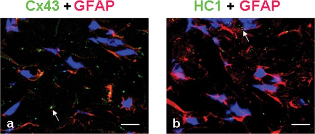
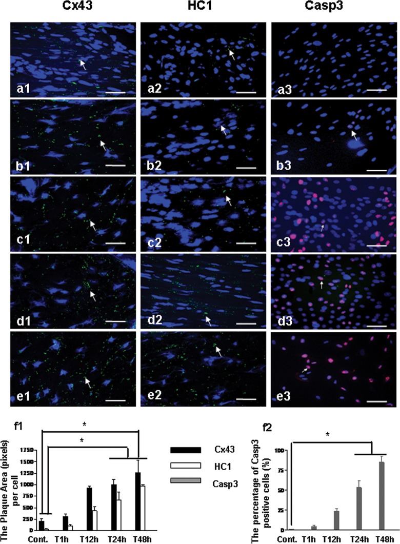
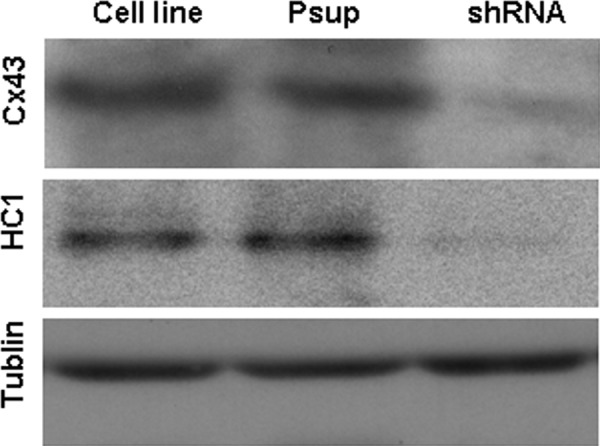
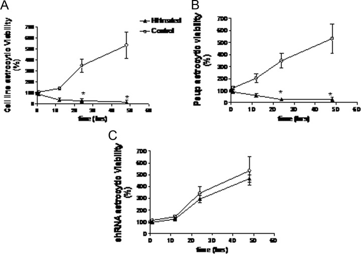
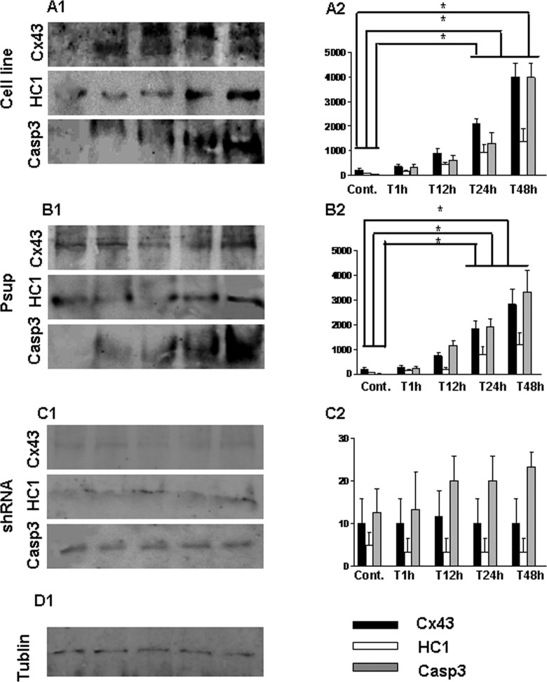
Wistar rat pups had the left common carotid artery cut, and they were exposed to 8% oxygen with free access to food and water until they were killed at 1, 12, 24, and 48 hours after the hypoxia-ischemia (HI) insult. Connexin 43 (Cx43), hemichannel (HC1), and caspase 3 (Casp3) in cerebral HI tissues were examined by immunohistochemistry and Western blot analyses. Astrocytes cell line, astrocytes transduced with a retroviral empty vector (Psup astrocyte), or a Cx43-specific small hairpin RNA (shRNA) construct (shRNA astrocytes) was treated with oxygen-glucose deprivation (OGD) insult. The viability of astrocytes was assessed by 3-(4,5-dimethylthiazol-2-yl)-2,5-diphenyltetrazolium bromide assay. The results showed the expression of Cx43, HC1, and Casp3 in rats' brain, and astrocytes and Psup astrocytes increased significantly after 24 hours of HI/OGD insult. Cell viability decreased after 24 hours of the insult. The results suggest that Cx43 and hemichannel are likely to mediate the astrocytic death after HI insult.

Keywords: oxygen–glucose deprivation (OGD); astrocytes; connexin 43; gap junction hemichannel; hypoxia–ischemia (HI); neuroprotection.


Conflict of interest statement

Declaration of Conflicting Interests: The authors declared no potential conflicts of interest with respect to the research, authorship, and/or publication of this article.

Figures

Figure 1.
Figure 1.
Double immunostaining for the expression of Cx43 + glial fibrillary acidic protein and HC1 + glial fibrillary acidic protein in the astrocytes from rat’s brain after HI insult. Cx43 and HC1 were stained green, glial fibrillary acidic protein was stained red, and nuclei were stained blue. The arrows show the positive staining of Cx43/HC1. Bar = 50 µm. Cx43 indicates connexin 43; GFAP, glial fibrillary acidic protein; HI, hypoxia–ischemia; HCl, hemichannel.
Figure 2.
Figure 2.
Immunostaining for the expression of Cx43, HC1, and Casp3 in rat’s brain after HI insult. Cx43, HC1, and Casp3 were stained green, and nuclei were stained blue. Bar = 25 µm. a1-3, The expression of Cx43, HC1, and Casp3 in SVZ from rat without the insult of HI (Cont.); b1-3, The expression of Cx43, HC1, and Casp3 in SVZ from rat 1 hour after HI (T1 hour); c1-3, The expression of Cx43, HC1, and Casp3 in SVZ from rat 12 hours after HI (T12 hour); d1-3, The expression of Cx43, HC1, and Casp3 from rat 24 hours after HI (T24 hour); e1-3, The expression of Cx43, HC1, and Casp3 in SVZ from rat 48 hours after HI (T48 hour); f1, The plaque area (pixels) of Cx43 and HC1 staining per cell with the time points. f2, The percentage of Casp3 positive cells (%) with the time points. *P < .05. Cx43 indicates connexin 43; Casp3, caspase 3; HCl, hemichannel; HI, hypoxia–ischemia; SVZ, subventricular zone.
Figure 3.
Figure 3.
Western blot analysis of Cx43, HC1, and Casp3 expression in subventricular zone of rat’s brain after HI insult. A, Protein expression of Cx43, HC1, Casp3, and tublin was identified from the tissue extracts in subventricular zone of rat’s brain. B, Bar histograms show the mean ± SD corresponding to data displayed in part A. *P < .05. Cx43 indicates connexin 43; Casp3, caspase 3; HCl, hemichannel; HI, hypoxia–ischemia; SD, standard deviation.
Figure 4.
Figure 4.
Western blot analysis to confirm the effect of transfection with null Cx43 gene shRNA in astrocytes. After 24 hours of the gene transfection, the lysate from astrocytes was resolved by SDS-PAGE, and immunopositive bands were semiquantified by scanning laser densitometry. Protein expression of Cx43, HC1, and tublin was identified, respectively. Cx43 indicates connexin 43; shRNA, small hairpin RNA; SDS-PAGE, sodium dodecyl sulfate–polyacrylamide gel electrophoresis.
Figure 5.
Figure 5.
Viability of astrocytes after OGD treatment. Astrocytes were cultured in 96-well plates at a density of 5000 cells/well and treated with OGD. Cells that did not undergo OGD treatment act as a control. Cell viability was assessed by the MTT assay (MTT measured in units of optical density at 570 nm). A, Cell line astrocytes; B, psup astrocytes; C, shRNA astrocytes. *P < .05. OGD indicates oxygen–glucose deprivation; MTT, 3-(4,5-dimethylthiazol-2-yl)-2,5-diphenyltetrazolium bromide; OGD, oxygen–glucose deprivation; shRNA, small hairpin RNA.
Figure 6.
Figure 6.
Western blot analysis of Cx43, HC1, and Casp3 expression from astrocytes following OGD treatment. Astrocytes were treated with OGD techniques. The lysates were resolved by SDS-PAGE, and immunopositive bands were semiquantified by scanning laser densitometry. Data are expressed as mean ± SD corresponding to data displayed, * P < .05. A1-2, Astrocytes cell line; B1-2, Psup astrocytes; C1-2, shRNA astrocytes; D1, Tublin was detected as a control. Cx43 indicates connexin 43; Casp3, caspase 3; HCl, hemichannel; OGD, oxygen–glucose deprivation; shRNA, small hairpin RNA; SDS-PAGE, sodium dodecyl sulfate–polyacrylamide gel electrophoresis; SD, standard deviation.

References

    1. Vannucci RC. Hypoxic-ischemic encephalopathy. Am J Perinatol. 2000;17(3):113–120. - PubMed
    1. Lai MC, Yang SN. Perinatal hypoxic-ischemic encephalopathy. J Biomed Biotechnol. 2011;2011:609813. - PMC - PubMed
    1. Vannucci RC, Perlman JM. Interventions for perinatal hypoxic-ischemic encephalopathy. Pediatrics. 1997;100(6):1004–1014. - PubMed
    1. Edwards AD, Brocklehurst P, Gunn AJ, et al. Neurological outcomes at 18 months of age after moderate hypothermia for perinatal hypoxic ischaemic encephalopathy: synthesis and metaanalysis of trial data. Br Med J. 2010;340:c363. - PMC - PubMed
    1. Cusato K, Bosco A, Rozental R, et al. Gap junctions mediate bystander cell death in developing retina. J Neurosci. 2003;23(16):6413–6422. - PMC - PubMed

LinkOut - more resources