. 2017 May 15:8:15406.
doi: 10.1038/ncomms15406.

Legionella effector Lpg1137 shuts down ER-mitochondria communication through cleavage of syntaxin 17

Affiliations

Legionella effector Lpg1137 shuts down ER-mitochondria communication through cleavage of syntaxin 17

Kohei Arasaki et al. Nat Commun. .

Abstract

During infection of macrophages, the pathogenic bacterium Legionella pneumophila secretes effector proteins that induce the conversion of the plasma membrane-derived vacuole into an endoplasmic reticulum (ER)-like replicative vacuole. These ER-like vacuoles are ultimately fused with the ER, where the pathogen replicates. Here we show that the L. pneumophila effector Lpg1137 is a serine protease that targets the mitochondria and their associated membranes. Lpg1137 binds to and cleaves syntaxin 17, a soluble N-ethylmaleimide-sensitive factor attachment protein receptor (SNARE) protein that is known to participate in the regulation of mitochondrial dynamics through interaction with the mitochondrial fission factor Drp1 in fed cells and in autophagy through interaction with Atg14L and other SNAREs in starved cells. Cleavage of syntaxin 17 inhibits not only autophagy but also staurosporine-induced apoptosis occurring in a Bax, Drp1-dependent manner. Thus, L. pneumophila can shut down ER-mitochondria communication through cleavage of syntaxin 17.


Conflict of interest statement

The authors declare no competing financial interests.

Figures

Figure 1
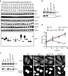
Figure 1. Stx17 is degraded on Legionella infection.
(a,b) HeLa-FcγRII cells were infected with (a) wild-type (WT) L. pneumophila or (b) one of the indicated strains at an MOI of 5. At 4 h after infection, cells were fixed and double stained with (a) an anti-Stx17 antibody and Hoechst 33342, or (b) an anti-Legoinella serum. Asterisks and arrows indicate infected cells and Legionella, respectively. Scale bar, 5 μm. (c) HeLa-FcγRII cells were infected with the WT Legionella or a dotA mutant strain at an MOI of 50. At the indicated times, cell lysates were prepared, and equal amounts of proteins were subjected to SDS–PAGE followed by IB with antibodies against calnexin (CNX) and Stx17. (d) THP1 cells were treated as described in a except for infection at an MOI of 25, and double stained with antibodies against Stx17 and Legoinella. Scale bar, 5 μm. (e) Similar experiments as in c except that the indicated strains were used for infection. Uncropped images of blots are shown in Supplementary Fig. 7.
Figure 2
Figure 2. Lpg1137 is responsible for the degradation of Stx17.
(a) HeLa-FcγRII cells were transfected with one of the indicated Lpg constructs. At 24 h after transfection, equal amounts of cell lysates were analysed by IB with the indicated antibodies. (b) Equal amounts of lysates from the indicated strains of Legionella were analysed by IB using antibodies against LidA and Lpg1137. (c) HeLa-FcγRII cells were infected with one of the indicated strains of Legionella at an MOI of 50 (left) or 5 (right). At 4 h after infection, cells were lysed and analysed by IB with antibodies against CNX and Stx17 (left) or double stained with an anti-Stx17 antibody and Hoechst 33342 (right). Asterisks and arrows indicate infected cells and Legionella, respectively. Scale bar, 5 μm. (d) Intracellular growth of Legionella and mutants. Host cells were infected with wild-type (WT) Legionella (blue line), Lpg1137 TM (black dot-line), Δ Lpg1137 (red line) or dotA mutant (black solid line). Total colony-forming units (CFU) of Legionella were determined at the indicated times as described in Methods. Values are means±s.d. (n=3). P<0.01 as compared with WT. No significant difference was observed between WT and Lpg1137 TM or Δ Lpg1137 mutant. Uncropped images of blots are shown in Supplementary Fig. 7.
Figure 3
Figure 3. Lpg1137 is a serine protease localized in MAM/mitochondria.
(a) HeLa-FcγRII cells were transfected with a plasmid encoding GFP (left) or GFP-Lpg1137 (right). At 24 h after transfection, cells were subjected to subcellular fractionation, and equal amounts of fractions were analysed by IB with the indicated antibodies. MS and MT denote microsomes and mitochondria, respectively. (b) HeLa-FcγRII cells were transfected with GFP-Lpg1137. At 4 h after transfection, DMSO (Vehicle), PMSF (1 mM) or MG132 (1 μM) was added to cells, and the cells were incubated for 20 h. Equal amounts of cell lysates were analysed by IB with the indicated antibodies. (c,d) HeLa-FcγRII cells were transfected with one of the indicated plasmids, and after 24 h equal amounts of lysates were analysed by IB with the (c) indicated antibodies. Alternatively, cells were fixed and stained with (d) an anti-Stx17 antibody. Scale bar, 5 μm. (e) His-Stx17 (0.2 μg) was incubated with MBP, MBP-Lpg1137 wild-type (WT) or MBP-Lpg1137 S68A (each 0.2 μg) for the indicated times at 37 °C. After incubation, samples were subjected to IB with antibodies against Stx17 and MBP. Uncropped images of blots are shown in Supplementary Fig. 7.
Figure 4
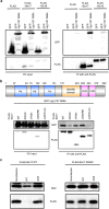
Figure 4. Stx17 interacts with Lpg1137 through the CHD+C region.
(a) HEK293-FcγRII cells were co-transfected with plasmids encoding GFP-Lpg1137 S68A and either FLAG, FLAG-Stx17 or FLAG-Stx18. At 24 h after transfection, cell lysates were prepared and immunoprecipitated with FLAG-M2 beads. The precipitated proteins were analysed by IB with antibodies against GFP and FLAG. (b) Schematic representation of Stx17 (upper panel). HEK293-FcγRII cells were co-transfected with plasmids encoding GFP-Lpg1137 S68A and FLAG-tagged full-length Stx17 or one of its mutants. Cell lysates were immunoprecipitated and analysed by IB with antibodies against GFP and FLAG (lower panel). (c) HeLa cells stably expressing FLAG-tagged wild-type (WT) Stx17 or the K254C mutant were transfected with a plasmid encoding non-tagged FcγRII. At 24 h after transfection, cells were non-infected or infected with WT Legionella or a dotA mutant at an MOI of 50 for 4 h. Equal amounts of cell lysates were analysed by IB with antibodies against CNX and FLAG. Uncropped images of blots are shown in Supplementary Fig. 7.
Figure 5
Figure 5. Lpg1137 eliminates the interactions of Stx17 with Atg14L and Drp1.
(a) HeLa-FcγRII cells expressing 3 × -FLAG-Atg14L (left) or HeLa-FcγRII cells (right) were non-infected or infected with Legionella at an MOI of 50 for 4 h. Before fixation, HeLa-FcγRII cells expressing 3 × -FLAG-Atg14L were incubated with EBSS for 2 h to induce autophagy. Cells were fixed and subjected to PLA using the indicated pairs, and the number of PLA dots was scored. Values are means±s.e.m (n=3). **P<0.001 and ***P<0.0001 as compared with noninfection. (b) HeLa-FcγRII cells were transfected with a plasmid encoding GFP, GFP-Lpg1137 wild-type (WT) or GFP-Lpg1137 S68A with or without 3 × -FLAG-Atg14L. At 24 h after transfection, cells expressing GFP constructs and 3 × -FLAG-Atg14L were incubated with EBSS for 2 h and fixed, or cells expressing only GFP constructs were fixed. PLA was performed using the indicated pairs. Values are means±s.e.m. (n=3). **P<0.001 and ***P<0.0001 as compared with GFP.
Figure 6
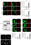
Figure 6. Ectopic expression of Lpg1137 blocks autophagosome formation.
(a) HeLa-FcγRII cells were transfected with a plasmid encoding GFP, GFP-Lpg1137 wild-type (WT) or GFP-Lpg1137 S68A. At 24 h after transfection, cells were incubated with EBSS (SV: left two columns) or EBSS plus 0.1 μM bafilomycin A1 (SV+bafilomycin A1, right two columns) for 2 h. After incubation, cells were fixed and stained with an anti-LC3 antibody. The bar graphs on the right show the ratio of the LC3 staining intensity of GFP-expressing to that of -nonexpressing cells. Values are means±s.d. (n=3). **P<0.001 as compared with GFP-Lgp1137 S68A. Scale bar, 5 μm. (b) HeLa-FcγRII cells were transfected with a plasmid encoding GFP or GFP-Lpg1137. At 24 h after transfection, cells were incubated with EBSS (SV)±0.1 μM bafilomycin A1 (Baf) for 2 h and then lysed. Equal amounts of the lysates were analysed by IB with the indicated antibodies. (c) HeLa-FcγRII cells expressing FLAG-DFCP1 (left two columns) or 3 × -FLAG-Atg14L (right two columns) were transfected with a plasmid encoding GFP, GFP-Lpg1137 WT or GFP-Lpg1137 S68A. At 24 h after transfection, cells were incubated with EBSS for 2 h, fixed and then stained with an anti-FALG antibody. The bar graphs shown at the bottom exhibit the ratio of the FLAG-DFCP1 or 3 × -FLAG-Atg14L staining intensity of GFP-expressing to that of -nonexpressing cells (50 cells each). Values are means±s.d. (n=3). **P<0.001 as compared with GFP-Lpg1137 S68A. Scale bar, 5 μm. (d) HeLa-FcγRII cells were transfected without (Mock) or with siRNA targeting Stx17 (Stx17 knockdown (KD)). At 48 h after transfection, cells were transfected with a plasmid encoding FLAG-DFCP1 or 3 × -FLAG-Atg14L, incubated for 24 h and then with EBSS for 2 h. The cells were fixed and stained with an anti-FLAG antibody. Scale bar, 5 μm. Uncropped images of blots are shown in Supplementary Fig. 7.
Figure 7
Figure 7. RavZ-positive puncta are not formed on Stx17 depletion.
(a) HeLa cells stably expressing Stx17 were transfected with a plasmid encoding GFP-RavZ (top and bottom rows) or both GFP-RavZ and mRFP-DFCP1 (middle row). At 24 h after transfection, cells were incubated with EBSS for 2 h, fixed and then stained with an antibody against LC3 (top low) or FLAG (bottom row). Arrows indicate representative GFP-RavZ-positive puncta. Scale bar, 5 μm. (b) HeLa-FcγRII cells were transfected without (Mock) or with siRNA (Stx17 knockdown (KD)) targeting Stx17. At 48 h after transfection, cells were transfected with plasmids encoding GFP-RavZ and mRFP-DFCP1. At 24 h after transfection, cells were incubated with EBSS for 2 h, fixed and observed by fluorescence microscopy. The numbers shown at the bottom of each panel indicate the intensity of GFP-RavZ or mRFP-DFCP1 staining in arbitrary unit (a.u.). Fifty cells expressing both GFP and mRFP were analysed in each experiment. Values are means±s.d. (n=3). P<0.01 (GFP-RavZ) or P<0.001 (mRFP-DFCP1). Scale bar, 5 μm.
Figure 8
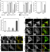
Figure 8. Loss of Stx17 suppresses STS-induced apoptosis.
(a) HeLa-FcγRII cells were transfected with one of the indicated plasmids. At 24 h after transfection, cells were mock-treated (Vehicle), or treated with 500 ng ml−1 TRAIL or 1 μM STS for 4 h and then fixed. Cells exhibiting nuclear condensation (left), cleaved caspase-3 (middle) and cytochrome c release from the mitochondria (right) were counted (100 GFP-expressing cells each). Values are means±s.d. (n=4). *P<0.01 and **P<0.001 as compared with GFP-Lpg1137 S68A. (b) HeLa-FcγRII cells with mock treatment or depleted (KD) of Stx17, Drp1 or Atg14L were treated with 1 μM STS for 4 h, fixed and then stained with an antibody against Bax (Supplementary Fig. 6). Notably, translocation of Bax to mitochondria increased the staining intensity in cells (Supplementary Fig. 6). One hundred cells were analysed in each experiment. The ratio of the Bax staining intensity between cells after and before STS treatment is plotted. Values are means±s.d. (n=4). ***P<0.0001 as compared with Mock. (c,d) HeLa-FcγRII cells were treated as described in b except for 2.5-h STS treatment to observe an early stage of apoptosis. Cells were fixed and double stained with the indicated antibodies. Scale bar, 5 μm. Insets in c are magnifications of boxed areas.

References

    1. Rowbotham T. J. Preliminary report on the pathogenicity of Legionella pneumophila for freshwater and soil amoebae. J. Clin. Pathol. 33, 1179–1183 (1980). - PMC - PubMed
    1. Horwitz M. A. & Silverstein S. C. Legionnaires' disease bacterium (Legionella pneumophila) multiples intracellularly in human monocytes. J. Clin. Invest. 66, 441–450 (1980). - PMC - PubMed
    1. Hubber A. & Roy C. R. Modulation of host cell function by Legionella pneumophila type IV effectors. Annu. Rev. Cell Dev. Biol. 26, 261–283 (2010). - PubMed
    1. Asrat S., de Jesús D. A., Hempstead A. D., Ramabhadran V. & Isberg R. R. Bacterial pathogen manipulation of host membrane trafficking. Annu. Rev. Cell Dev. Biol. 30, 79–109 (2014). - PubMed
    1. Horwitz M. A. The Legionnaires' disease bacterium (Legionella pneumophila) inhibits phagosome–lysosome fusion in human monocytes. J. Exp. Med. 158, 2108–2126 (1983). - PMC - PubMed

Publication types